Fig. 4
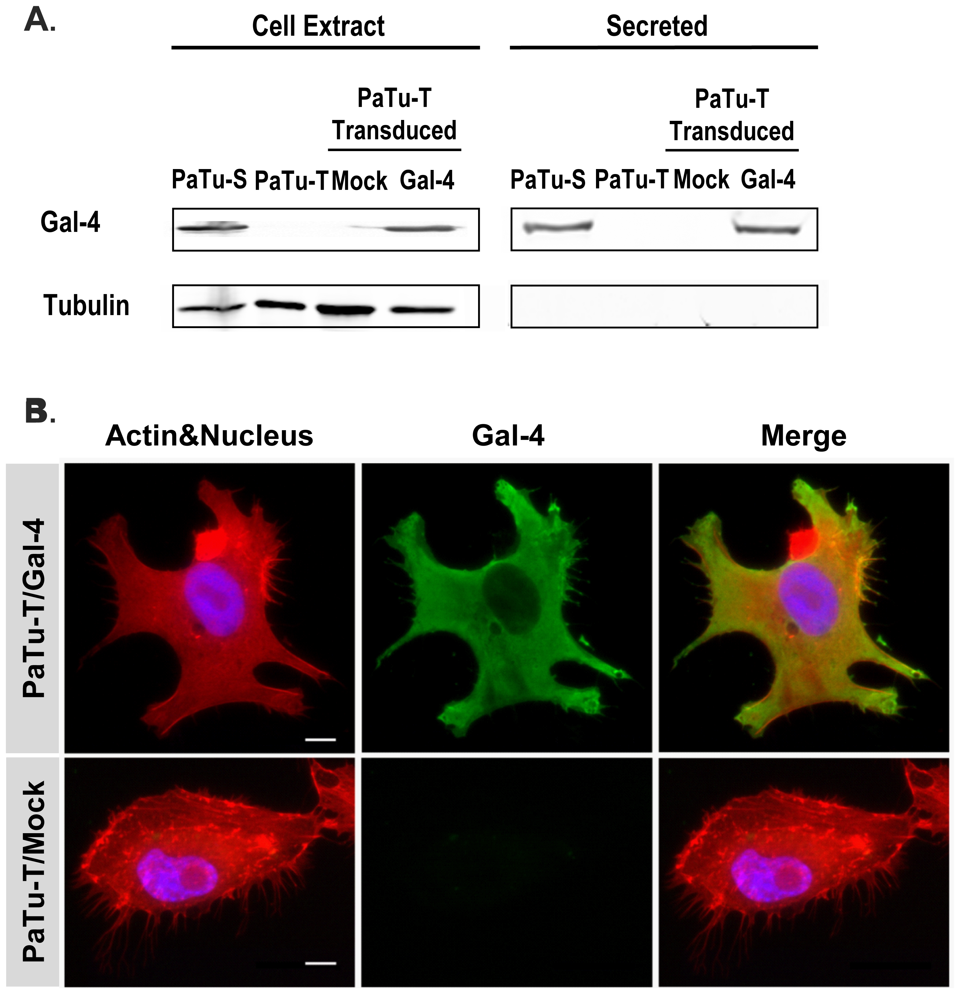
Gal-4 protein levels in PaTu-S and PaTu-T cells, and localization of Gal-4 in PaTu-T/Gal-4.
A) Proteins from whole-cell extracts (75 ug total protein) and culture medium (4 days culture, 25 ul) of PaTu-S (P-S), PaTu-T (P-T), PaTu-T/Gal-4 (P-T/Gal-4) and PaTu-T/mock (P-T/M) were separated by SDS-PAGE. After transfer of the proteins to a nitrocellulose membrane, the blots were stained using goat anti-hGal-4 for detection of Gal-4, and mouse anti-tubulin as control for the presence of intracellular protein. B) Photographs of representative ICC analysis of the cellular localization of Gal-4 in PaTu-T/Gal-4 and PaTu-T/mock cells. Gal-4 was detected using Alexa-labeled anti-Gal-4 Abs (green), Actin was stained using Phalloidin (red) and nucleus staining obtained using HOESCHS (blue); the third panel shows the merging of the different stainings. Bar = 25 μm.

