
Fig. 4
- ID
- ZDB-IMAGE-130826-47
- Publication
- Chapman et al., 2013 - Axonal Transport Defects in a Mitofusin 2 Loss of Function Model of Charcot-Marie-Tooth Disease in Zebrafish
- All Figures
- Figures for Chapman et al., 2013
|
Fig. 4
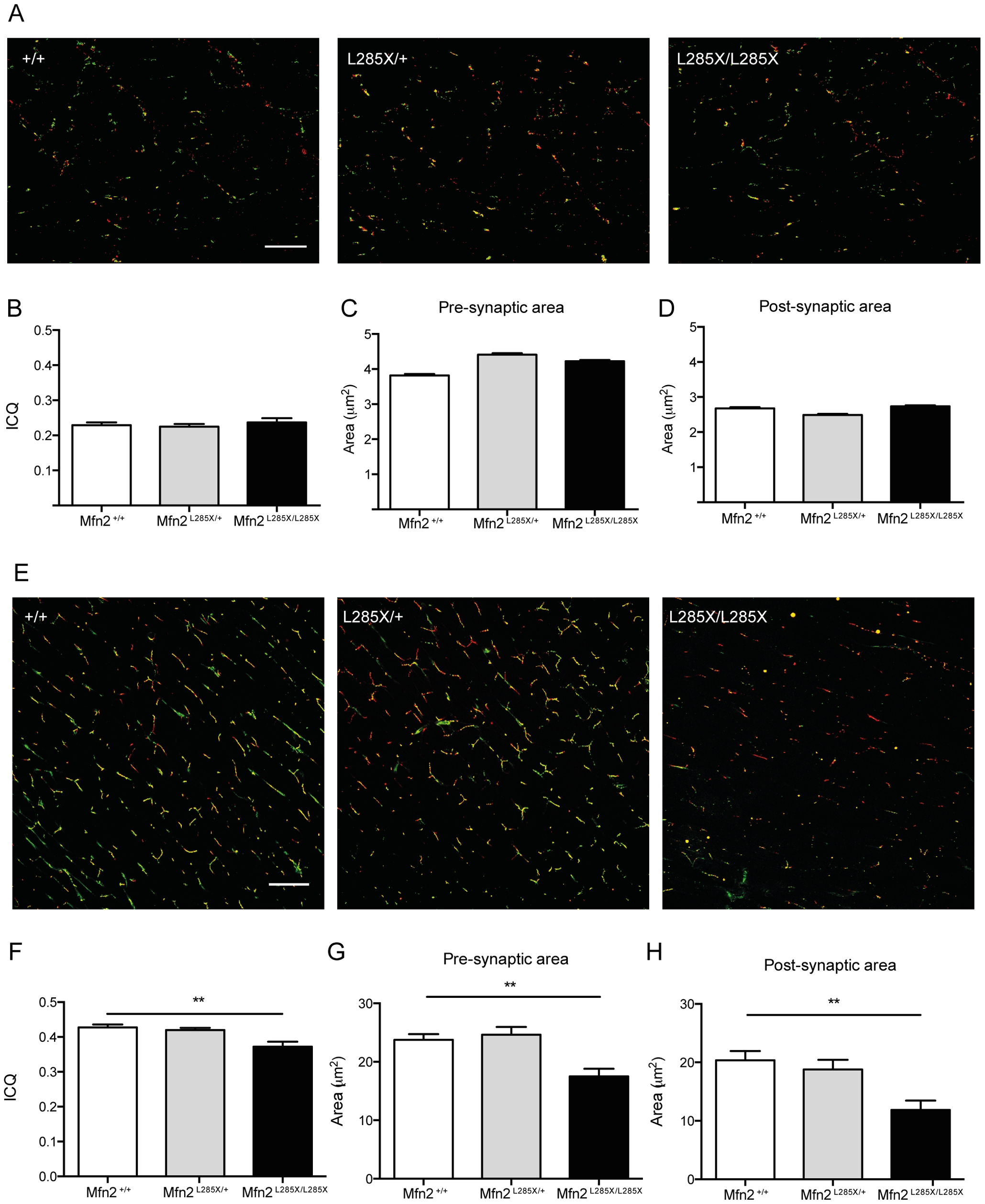
Alterations of NMJ pathology in MFN2L285X larvae (A–D) and adults (E–H).
(A) Representative images of dual immunofluorescence staining of whole mount 15d larvae of each genotype for α-Bungarotoxin (green) and SV2 (red). (B) ICQ analysis of 3 larvae per group reveals no alteration of SV2/α-Bungarotoxin co-localisation in MFN2L285X/+ or MFN2L285X/L285X larvae. Compared to siblings the (C) pre-synaptic area, and (D) post-synaptic area are not significantly different in MFN2L285X/+ or MFN2L285X/L285X larvae. (E) Representative images of a-Bungarotoxin (green) and SV2 (red) in 200 day-old zebrafish. (F) ICQ is significantly reduced in MFN2L285X/L285X at this stage, and there are significant reductions in the pre- and post-synaptic area (G and H). Scale bars = 10 μm.