
Fig. 4
- ID
- ZDB-IMAGE-130826-34
- Publication
- Huang et al., 2013 - Igf Signaling is Required for Cardiomyocyte Proliferation during Zebrafish Heart Development and Regeneration
- All Figures
- Figures for Huang et al., 2013
|
Fig. 4
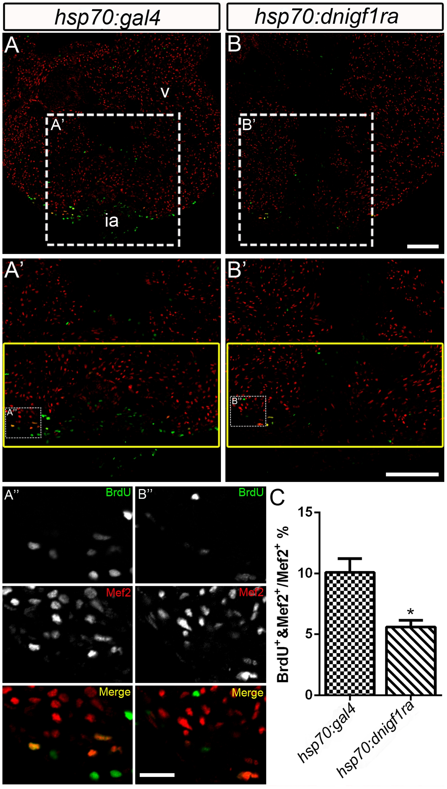
Igf signaling is required for cardiomyocyte proliferation during zebrafish heart regeneration.
Cardiomyocyte proliferation was decreased after Igf signaling was blocked during zebrafish heart regeneration. Tg(hsp70:gal4) control (n = 5) (A, A′, A′′) and Tg(hsp70:dnigf1ra-GFP) transgenic zebrafish (n = 7) (B, B′,B′′) zebrafish were heat shocked for 1 h at 38°C after amputation from 2–10 dpa. BrdU (green) and Mef2 (red) double positive cells indicate proliferating cardiomyocytes (A, A′, B, B′). A′ and B′ are the higher magnification images of the dashed boxes in A and B. A′′ and B′′ are the higher magnification images of the dashed boxes in A′ and B′. The yellow box indicates the wound area; cardiomyocytes were counted in this region. (A′′ and B′′), BrdU (green) and Mef2 (red) staining were shown as separated channel images (black and white) or merged color images. ia: injured area, v: ventricle. Scale Bar: (B, B′) = 100 μm, (B′′) = 20 μm. (C) Quantification of BrdU positive cardiomyocytes (Mef2 positive) ± S.E. A significant decrease (*p<0.05) in cardiomyocyte proliferation was detected in Tg(hsp:dnigf1ra-GFP) fish.