Fig. 1
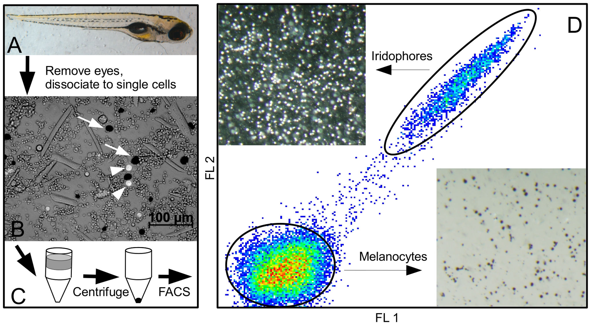
Purification procedure for melanocytes, iridophores, and retinal pigmented epithelium.
Zebrafish are grown to the desired time point; shown in (A) is a six day old fish. (B) Fish are dissociated to a single cell suspension; black melanocytes (arrows) and reflective iridophores (arrowheads) are visible as a small percentage of all cells. (C) Cells are placed atop a Percoll density gradient and centrifuged. (D) The resulting cell pellet is resuspended and analyzed by FACS. Shown is a characteristic FACS plot demonstrating the relative positions of melanocyte and iridophore gates (ovals). Sorted iridophores are shown on the upper left of (D) under incident light. Sorted melanocytes are on the lower right using trans-illumination.

