
Fig. 4
- ID
- ZDB-IMAGE-130822-4
- Publication
- Sah et al., 2013 - Ion channel-kinase TRPM7 is required for maintaining cardiac automaticity
- All Figures
- Figures for Sah et al., 2013
|
Fig. 4
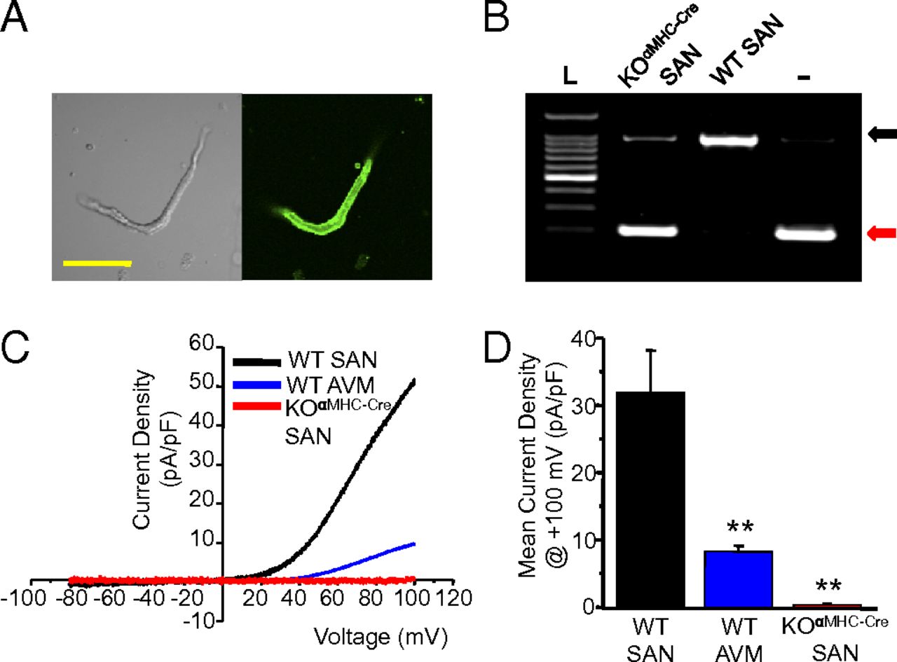
TRPM7 is highly expressed in murine SAN. (A) Confocal images of an isolated SAN freshly dissociated from adult αMHC-Cre-ROSA26mTmG heart: differential interference contrast (Left) and fluorescence GFP image (Right). (Scale bar, 50 μm.) (B) PCR across exon 17 from genomic DNA isolated from dissected KOαMHC-Cre and WT SAN. Tail DNA from Trpm7fl/- (-) serves as a positive control for Trpm7 exon 17 deletion. Black arrow, full-length exon 17. Red arrow, deleted exon 17. (C) Representative TRPM7 current-voltage traces measured in WT SAN, WT AVM, and KOαMHC-Cre SAN. (D) Mean TRPM7 current density in WT SAN (ITrpm7, WT SAN = 32.0 ± 6.2 pA/pF, n = 6), WT AVM (ITrpm7, AVM = 8.3 ± 0.9 pA/pF, n = 5), and SAN (ITrpm7, KOαMHC-Cre SAN = 0.4 ± 0.2 pA/pF, n = 4, P < 0.01).