
|
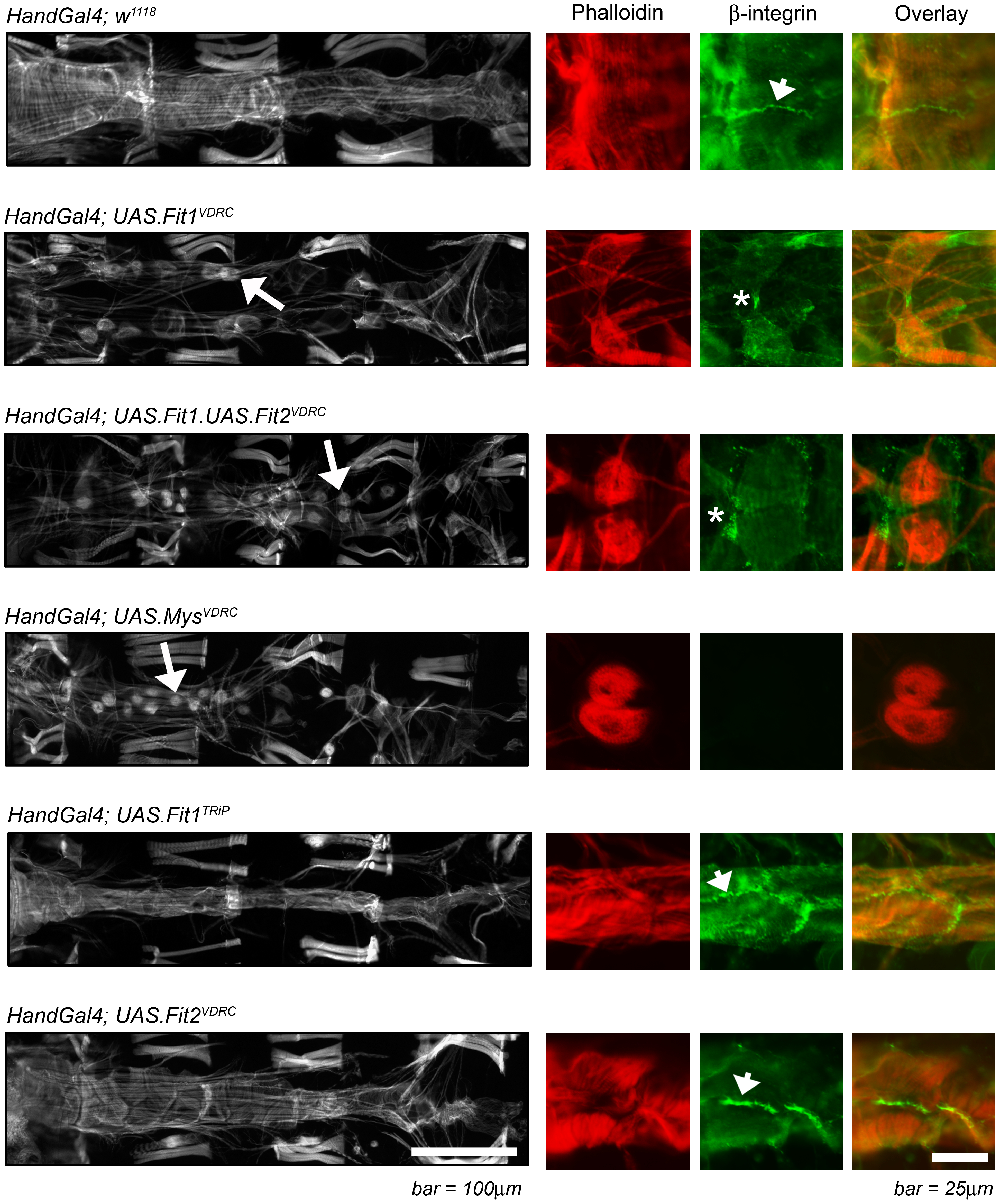
Fig. 2
The monochrome micrographs show the adult Drosophila heart (segments A2 to A5) stained with phalloidin, oriented with the anterior region to the left. Genes were silenced using the Hand-Gal4 enhancer which drives expression in cardiomyocytes (as well as pericardial nephrocytes and enterocytes of the gut) and abnormal cardiomyocyte/heart morphology is highlighted by the arrows. The colour panels show higher magnification micrographs of at least two cardiomyocytes stained with phalloidin (red) and antibodies to the Drosophila β-integrin, myospheroid (green); arrowheads indicate normal integrin staining between cardiomyocytes; asterisks indicate an abnormal staining pattern associated with loss of cardiomyocyte junction integrity. A wild type phenotype (Hand-Gal4; w1118), is characterised by contiguous cardiomyocytes and β-integrin staining between cardiomyocytes.