
Fig. S2
- ID
- ZDB-IMAGE-130813-8
- Publication
- Reimer et al., 2013 - Dopamine from the Brain Promotes Spinal Motor Neuron Generation during Development and Adult Regeneration
- All Figures
- Figures for Reimer et al., 2013
|
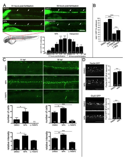
Fig. S2 (related to Fig. 2) Dopaminergics influence development of Islet- 1:GFP+ motor neurons, but not Pax2a+ or Glyt2:GFP+ interneurons. A: Lateral views of the trunk region of living islet-1:GFP transgenic animals under a stereo-microscope are shown. The dopamine agonist NPA (drug exposure 24 à 51 hpf; ANOVA, ***P < 0.001 compared to DMSO) significantly increases and the antagonist Haloperidol (drug exposure 24 to 56 hpf; ANOVA, **P < 0.01 compared to DMSO) decreases the percentage of embryos with islet- 1:GFP+ motor neurons (arrows) in a dose dependent fashion. B: The D4 specific antagonist L-745870 attenuates development of islet-1:GFP+ motor neurons, which cannot be overcome by the addition of NPA (56 hpf; ANOVA, ***P < 0.0001). C: Visual scoring of islet-1:GFP embryos is confirmed by cell counts and fluorescence intensity measurements. Lateral views of z-projected confocal image stacks of the mid trunk region are shown. At 51 hpf (left column), cell number (*P < 0.05; Mann-Whitney U-test, one tailed) and fluorescence intensity are significantly increased (*P < 0.05; Mann-Whitney Utest, one tailed) in NPA treated embryos. At 56 hpf (right column), cell number (ANOVA, ***P < 0.0001) and fluorescence intensity (ANOVA, ***P < 0.0001) are significantly lower in L-745870 treated embryos and still increased in NPA treated animals compared to DMSO controls (ANOVA, *P < 0.05; **P < 0.01). Fluorescence intensity of an expression domain in the cerebellum was unchanged by any of the treatments (data not shown). D: Lateral trunk views of embryos at 33 hpf are shown (drug exposure 24 to 33 hpf; genotypes and drugs are indicated). NPA has no effect on the numbers of glyt2:GFP+ or pax2a:GFP+ interneurons. Error bars represent SEM. Scale bars: A: 100 μm; C: 60 μm, D: 50 μm.
Reprinted from Developmental Cell, 25(5), Reimer, M.M., Norris, A., Ohnmacht, J., Patani, R., Zhong, Z., Dias, T.B., Kuscha, V., Scott, A.L., Chen, Y.C., Rozov, S., Frazer, S.L., Wyatt, C., Higashijima, S., Patton, E.E., Panula, P., Chandran, S., Becker, T., and Becker, C.G., Dopamine from the Brain Promotes Spinal Motor Neuron Generation during Development and Adult Regeneration, 478-491, Copyright (2013) with permission from Elsevier. Full text @ Dev. Cell