
Image
|
Figure Caption
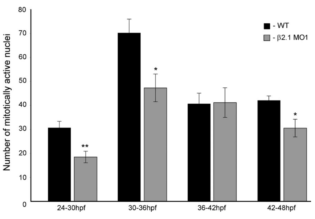
Fig. 8 Bromodeoxyuridine (BrdU) assay of cardiomyocyte cells undergoing DNA replication, spanning 6-hr periods of cardiac development. β2.1-depleted embryos display a significantly reduced rate of mitosis (asterisks) at 24–30 hpf (P = 0.002), 30–36 hpf (P = 0.01), and 42–48 hpf (P = 0.01). n = 10 wild-type and 10 morphant embryos per time period.
Figure Data
Acknowledgments
This image is the copyrighted work of the attributed author or publisher, and
ZFIN has permission only to display this image to its users.
Additional permissions should be obtained from the applicable author or publisher of the image.
Full text @ Dev. Dyn.