
Fig. 6
- ID
- ZDB-IMAGE-130808-66
- Publication
- Duval et al., 2013 - Longitudinal fluorescent observation of retinal degeneration and regeneration in zebrafish using fundus lens imaging
- All Figures
- Figures for Duval et al., 2013
|
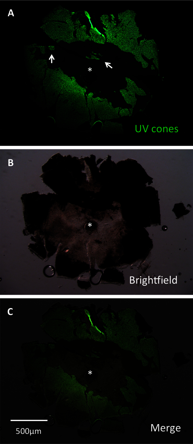
Fig. 6
A lesioned retina was dissected to assess efficacy of lesioning methods. This retina was dissected at 3 days post-lesion (DPL) and flatmounted for imaging. Lesioning occurred across the eye. Lesion edges were observed to vary between clean, straight edges (center of retina) and variable shapes (left and right edges, representing periphery of retina). Patches of surviving cones (arrows) were commonly observed. A: The image is presented showing green fluorescent protein (GFP), which is contained in ultraviolet-sensitive (UV) cones. B: A brightfield view of the same retina. C: A merge of images A and B is presented. The asterisk indicates the optic nerve head (ONH).