
Fig. S4
- ID
- ZDB-IMAGE-130808-59
- Publication
- Lee et al., 2013 - An exclusively mesodermal origin of fin mesenchyme demonstrates that zebrafish trunk neural crest does not generate ectomesenchyme
- All Figures
- Figures for Lee et al., 2013
|
Fig. S4
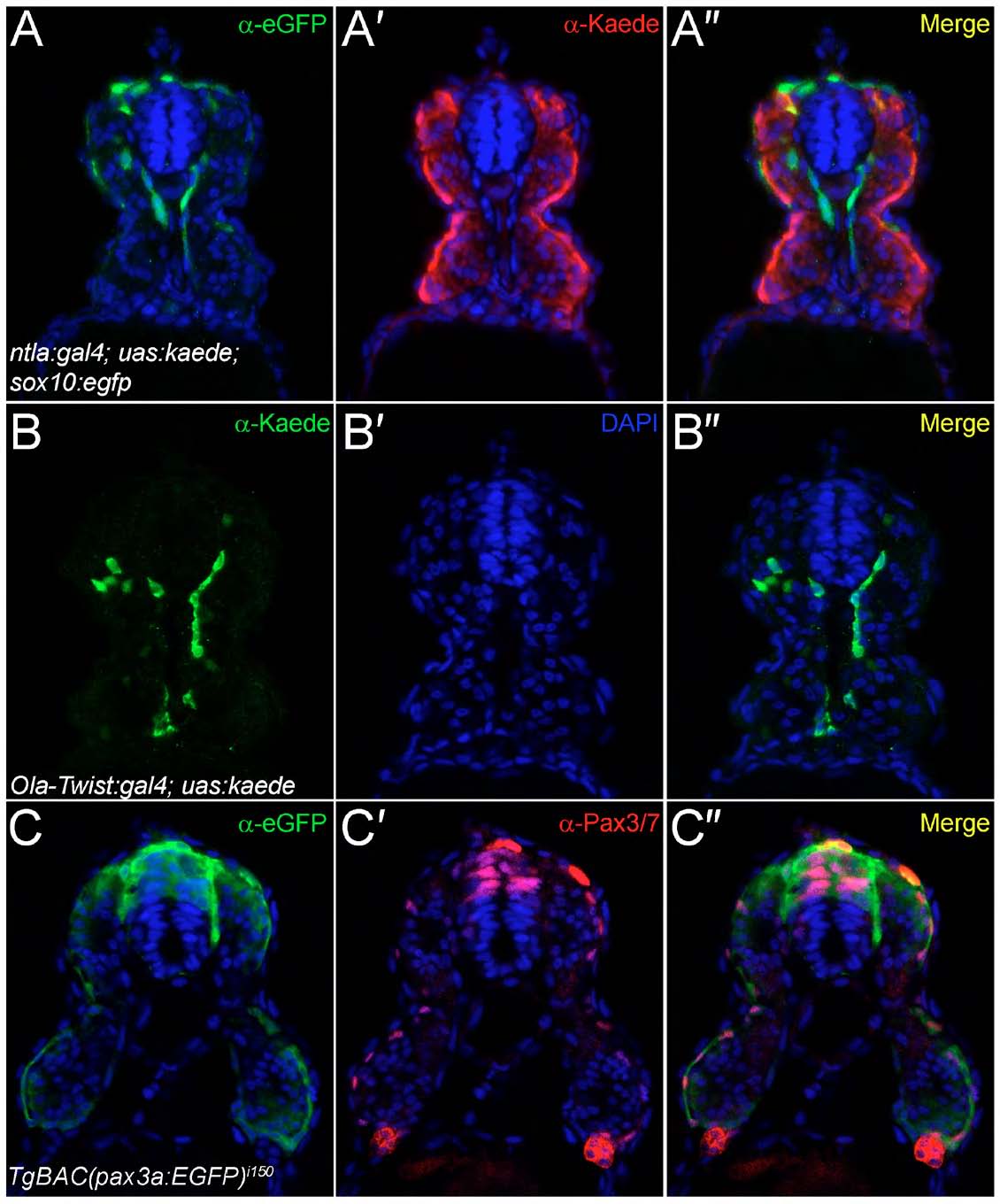
Analysis of transgenic lines used to define the origin of fin mesenchyme. Transverse cryosections of the trunk region of the ntla:gal4; uas:kaede (A-A"), the Ola-Twist:gal4; uas:kaede (B-B") and the TgBAC(pax3a:EGFP)i150 (C-C") transgenic lines at 24 hpf, imaged by confocal microscopy following fluorescent immunostaining with antibodies against eGFP (A-A",C-C"), Kaede (A-A",B-B") and Pax3/7 (C-C0). All sections were counterstained with DAPI (blue). (A-A") To demonstrate exclusion of Kaede expression from neural crest, the ntla:gal4; uas:kaede line was crossed to the sox10:egfpba2 line and immunostained for eGFP (A) and Kaede (A′). Restriction of Kaede to the mesoderm and exclusion from the neural crest can be seen in the superimposed image (A"). (B-B") Expression of Kaede in the Ola-Twist:gal4; uas:kaede line is largely restricted to the sclerotomal compartment of the somites as seen by immunostaining for Kaede expression (B), which can be seen in a medial somitic location (B") and far removed from the superficial dermomyotome domain. Occasional myotome expression can be observed in this line (B"). (C-C") The TgBAC(pax3a:EGFP)i150 line faithfully recapitulates Pax3 expression in the dermomyotome as shown by comparing eGFP immunofluorescence (C) with Pax3/7 immunoreactivity (C′). By superimposition of the two confocal images, eGFP-positive dermomyotome cells at the somite surface have Pax3/7-positive nuclei, with strong eGFP and Pax3/7 colabelling in the dorsal neural tube and neural crest also apparent (C").