Fig. S3
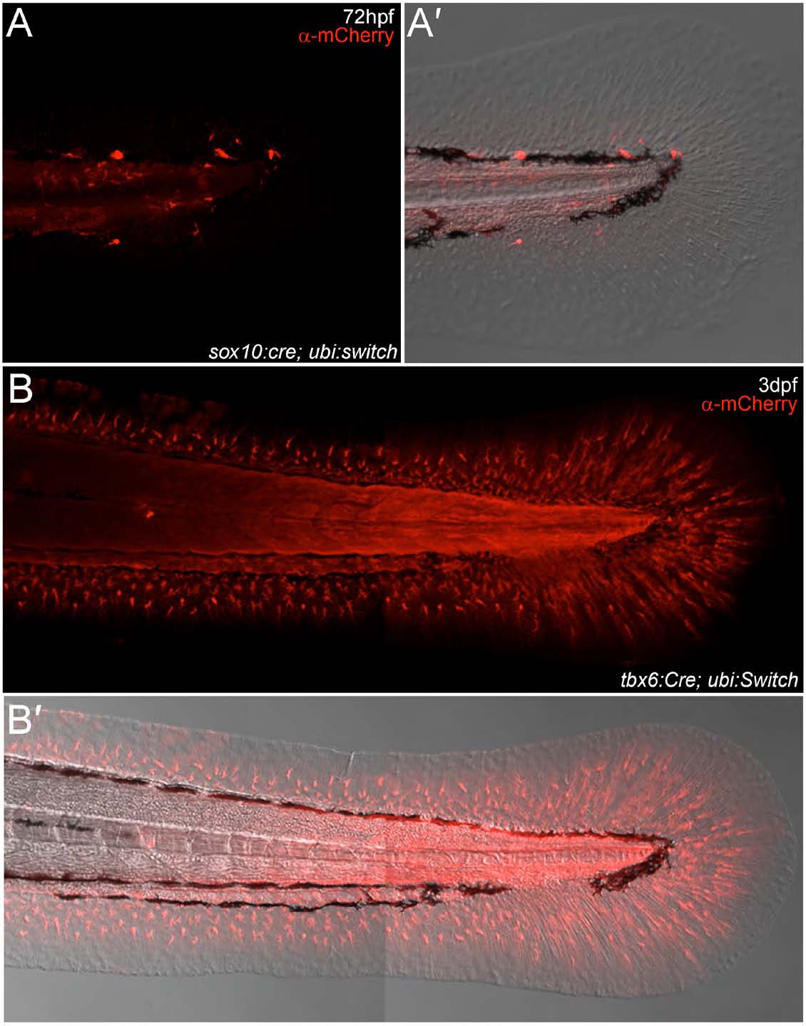
Permanent lineage analysis confirms the origin of embryonic fin mesenchyme. (A,A′) Lateral confocal images of 72-hpf sox10:Cre; ubi:switch embryos fluorescently immunostained with an antibody detecting mCherry. Image of mCherry expression within the posterior trunk and fin (A) and also superimposed on a Nomarski view to show limited cells within the fin (A′). These embryos have neural crest lineages permanently labelled with mCherry, and show labelled cells within the trunk in locations and with morphology consistent with described neural crest derivatives. Fin mesenchyme is unlabelled. (B,B′) Lateral confocal images of 3-dpf tbx6:Cre; ubi:switch embryos fluorescently immunostained with an antibody detecting mCherry. (B) mCherry expression within the posterior trunk and fin with widespread expression visible in the fin mesenchyme and muscle of trunk. (B′) Fluorescent image superimposed on a Nomarski view outlining expression domains within the fin.

