
Fig. 2
- ID
- ZDB-IMAGE-130808-51
- Genes
- Publication
- Lee et al., 2013 - An exclusively mesodermal origin of fin mesenchyme demonstrates that zebrafish trunk neural crest does not generate ectomesenchyme
- All Figures
- Figures for Lee et al., 2013
|
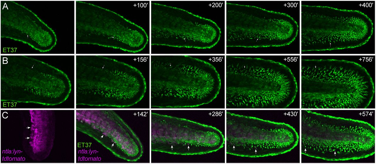
Fig. 2
Fin mesenchyme cells emerge from trunk mesoderm. Stills of time-lapse Movies 1-3 (see supplementary material Movies 1-3) of the tail of the ET37 line alone (A,B) or crossed to the ntla:lyn-tdtomato transgenic line (C). eGFP is shown in green and membrane-tdTomato is in magenta. Left panels are taken at <26 hpf (A), 29 hpf (B) and 22 hpf (C), with subsequent time points (indicated in minutes) shown in panels to the right. Examples of fin mesenchyme cells are indicated (arrows) emerging from the ventral (A,C) and dorsal (B) mesoderm into the adjacent fin. First panel of C is shown without eGFP signal to highlight the epithelial nature of the cells within the mesoderm prior to fin immigration.