
IMAGE
Fig. 4
- ID
- ZDB-IMAGE-130808-13
- Antibodies
- Publication
- Hayes et al., 2013 - Expression of glycosaminoglycan epitopes during zebrafish skeletogenesis
- All Figures
- Figures for Hayes et al., 2013
Image
|
Figure Caption
Fig. 4
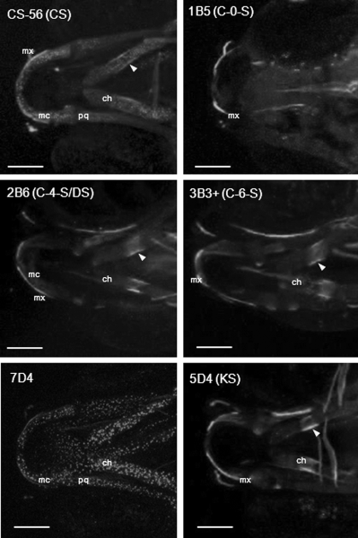
Chondroitin and keratan sulfate expression in the trunk and developing vertebral column. Immunohistochemistry of distinct chondroitin and keratan sulfate moities (as labelled) at 4 dpf and 8 dpf. All are lateral views with anterior to left. Trunk images at 4 dpf show a 4-somite span at the level of the cloaca. At 8 dpf, the images show an 8–9-somite span from the anterior end of the fish. Scale bars = 100 μm in all panels. hm, horizontal myoseptum; sb, somite boundary; sk, skin; nc, notochord; ha, haemal arch; na, neural arch; vb, vertebral body.
Figure Data
Acknowledgments
This image is the copyrighted work of the attributed author or publisher, and
ZFIN has permission only to display this image to its users.
Additional permissions should be obtained from the applicable author or publisher of the image.
Full text @ Dev. Dyn.