
Fig. 2
- ID
- ZDB-IMAGE-130807-2
- Publication
- Fang et al., 2013 - Characterization of transgenic zebrafish lines that express GFP in the retina, pineal gland, olfactory bulb, hatching gland, and optic tectum
- All Figures
- Figures for Fang et al., 2013
|
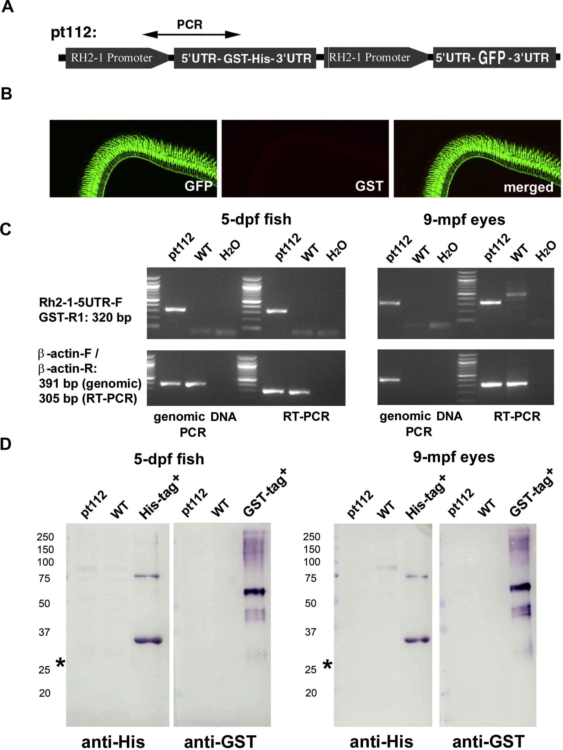
Fig. 2 Transgenic zebrafish line Tg(LCRRH2-RH2-1:GFP)pt112, expresses GFP but not GST-His fusion in the retina. (A) The schematic illustrates the structures and tandem arrangement of the GST-His and GFP reporter genes in the transgene construct that was used to generate the Tg(LCRRH2-RH2-1:GFP)pt112 line. The regions amplified by PCR genotyping reactions are indicated with double-headed arrows. (B) In the adult Tg(LCRRH2-RH2-1:GFP)pt112 retina at 8 mpf (months postfertilization), the expression of the GST-His protein was undetectable, whereas GFP expression is very strong. (C) RT-PCR and genomic DNA PCR analyses demonstrated that the GST-His gene was present in the Tg(LCRRH2-RH2-1:GFP)pt112 genome, and was apparently transcribed at both 5 dpf and 9 mpf. (D) Western blotting analyses confirmed that GST-His fusion protein was not expressed in Tg(LCRRH2-RH2-1:GFP)pt112 fish at both 5 dpf and 9 mpf. The asterisks indicate the position where the fusion protein was supposed to be. Unrelated His-Tagged and GST-tagged recombinant proteins expressed in Escherichia coli were used as controls for Western blotting conditions.
Reprinted from Gene expression patterns : GEP, 13(5-6), Fang, W., Bonaffini, S., Zou, J., Wang, X., Zhang, C., Tsujimura, T., Kawamura, S., and Wei, X., Characterization of transgenic zebrafish lines that express GFP in the retina, pineal gland, olfactory bulb, hatching gland, and optic tectum, 150-9, Copyright (2013) with permission from Elsevier. Full text @ Gene Expr. Patterns