
|
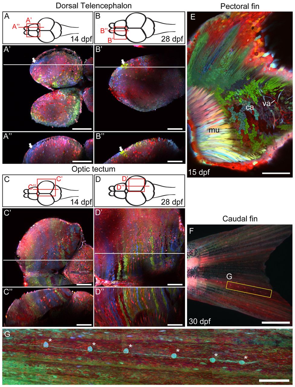
Fig. 8 Zebrabow labeling in different organ systems. (A-D′) Two-photon image of the dorsal telencephalon and optic tectum at late larval stage (14 dpf, A-A′ and C-C′) and juvenile stage (28 dpf, B-B′ and D-D′). A-D show diagrams of the zebrafish brain, viewed dorsally. A2-D2 show horizontal optical sections of the areas highlighted in the diagrams above. A′-D′ show sagittal optical sections along the horizontal lines in the middle panel. Clusters of single-colored cells are seen at different regions of the dorsal telencephalon (white arrows). (E) The pectoral fin of a 15 dpf animal, viewed from the side. Muscle (mu), cartilage (ca) and vasculature (va) can be seen in this view. (F,G) In the caudal fin, an array of lateral line organs (asterisks) extend from the base of the tail to the edge of the caudal fin (G). Scale bars: 100 μm in A-E; 1 mm in F; 250 μm in G.