
Fig. S1
- ID
- ZDB-IMAGE-130802-35
- Publication
- Choudhuri et al., 2013 - Translation initiation factor eIF3h targets specific transcripts to polysomes during embryogenesis
- All Figures
- Figures for Choudhuri et al., 2013
|
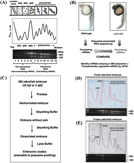
Fig. S1 Polysome profile analysis and the strategy of subsequent RNA-seq of polysome-associated mRNAs in zebrafish embryos. (A) A cell-free extract prepared from actively translating cells is loaded onto a linear 10–50% sucrose gradient and subjected to velocity gradient centrifugation. Free 40S, 60S, and 80S ribosomes, in addition to polysomes, are size fractionated from the translationally inactive mRNAs and messenger ribonuclear protein (MRNP) particles. The A254 profile is analyzed with an attached UV-absorbance monitor. The position and integrity of the ribosomal components in the separated fractions are demonstrated by isolating total RNA from individual fractions and analyzing aliquots by gel electrophoresis. The presence of 18S, 28S, or equimolar 18S and 28S ribosomal RNAs (rRNAs) identifies the positions of 40S, 60S, and 80S ribosomes, respectively. Polysomal fractions also yield both 18S and 28S rRNAs and consist of translating mRNAs. (B) To identify genes that are translationally regulated, stage-matched WT and morphant embryos were collected, and whole-cell lysates were processed as in A. Both total RNA and polysome-associated RNA were used to generate cDNA libraries that were analyzed by deep sequencing to identify transcripts underrepresented in the morphant samples (in the polysome, but not the total RNA samples). Here we compared eif3ha and control samples at 24 hpf. (C) Flowchart indicating the protocol optimized to obtain reproducible polysome profiles from cell-free extracts of zebrafish embryos. (D) A typical polysome profile prepared from fresh (nonfrozen) WT embryos. (E) A representative polysome profile prepared from frozen WT embryos. The positions of 40S, 60S, and 80S ribosomes are indicated.