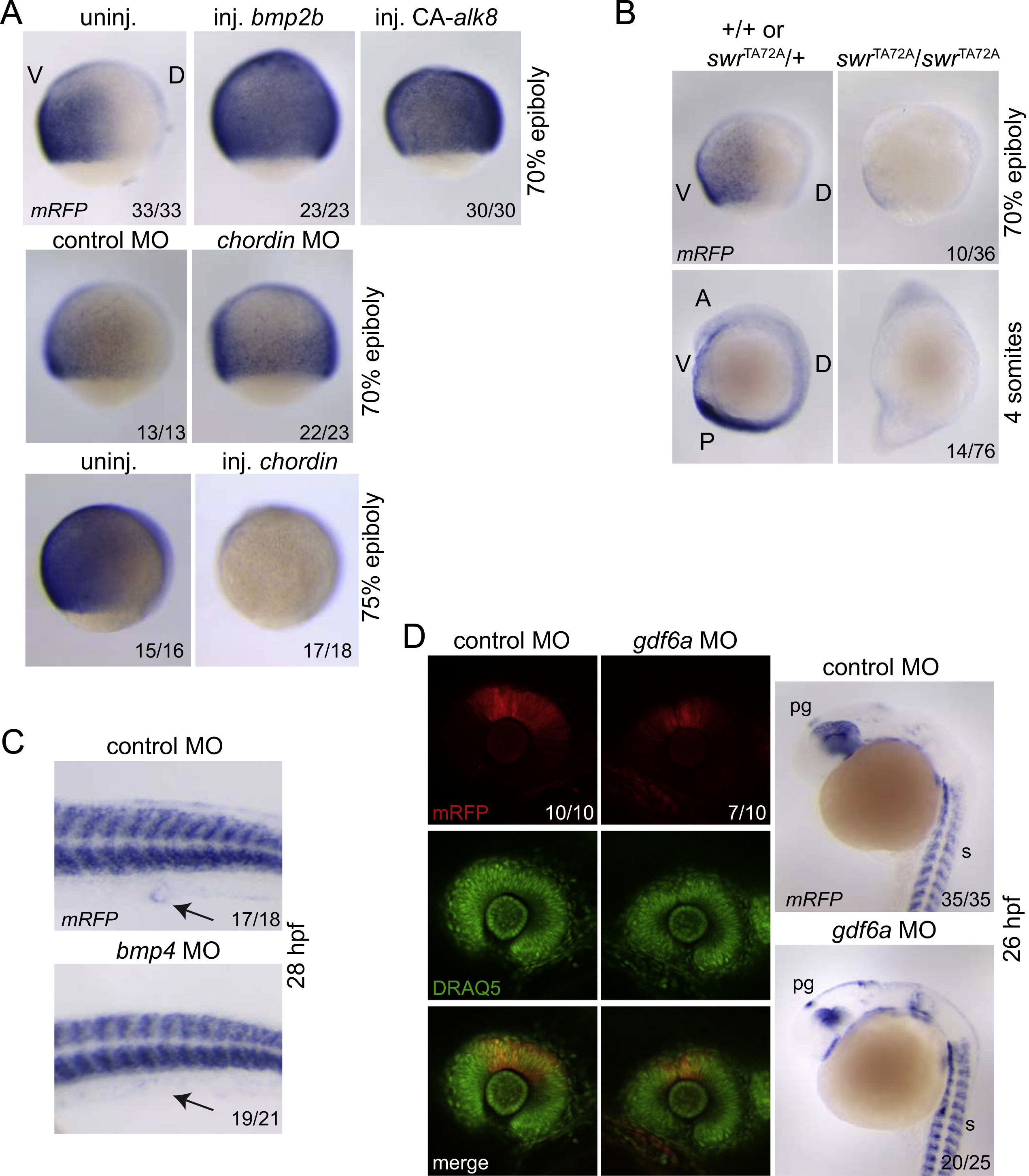
Fig. 2 mRFP expression in BRE-mRFPembryos is abona fidereadout for BMP and GDF activities (A) mRFP ISH in BRE-mRFP embryos injected with chordin, bmp2b or CA-alk8 mRNAs or chordin MO. (B) BRE-mRFP expression in swrTA72A/swrTA72A homozygous embryos compared with +/+ or swrTA72A/+ siblings. The embryos shown resulted from a cross between swrTA72A/+ heterozygotes that contained the BRE-mRFP transgene. (C) mRFP ISH in BRE-mRFP embryos injected with a control or bmp4 MO. Arrows point to the cloaca. (D) BRE-mRFP embryos were injected with a control or gdf6a MO and mRFP expression in the dorsal retina was analysed (fluorescence and ISH). pg, pineal gland; s, somites. In (A) and (B), lateral views are shown; V, ventral; D, dorsal; A, anterior; P, posterior. In all cases, stages are indicated and the number of embryos out of the total analysed that showed the presented staining pattern is given. For (B), this number represents the number of homozygous mutant embryos out of the total number of embryos analysed.
Reprinted from Developmental Biology, 378(2), Ramel, M.C., and Hill, C.S., The ventral to dorsal BMP activity gradient in the early zebrafish embryo is determined by graded expression of BMP ligands, 170-82, Copyright (2013) with permission from Elsevier. Full text @ Dev. Biol.

