
|
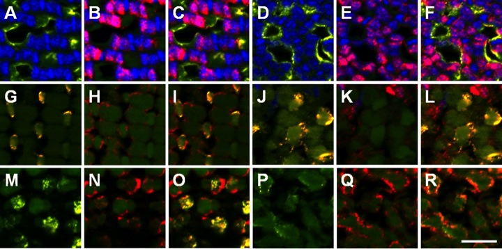
Fig. 2 Cone subtypes can be identified by the vertical tiering distribution. A–F: UV opsin (yellow) marks UV-sensitive cones, and zpr-1 staining (red) marks the cell bodies of the red-/green-sensitive double cones in the adult mosaic (A-C) and the larval retina (D-F) at the vertical location of the basal body. G-L: Blue opsin (yellow) labels blue-sensitive cones, and zpr-1 staining (red) labels red-/green-double cones in the adult mosaic (G-I) and the larval retina (J-L) at the vertical location of the basal bodies. M-R: Green cone opsin (yellow) labels green-sensitive cones and colocalizes with one member of the double-cone pair labeled by zpr-1 staining (red) in the adult mosaic (M-O) and the larval retina (P–R). A magenta-green version of this figure is provided in the Supporting Information. Scale bar = 10 μm.