
Fig. S4
- ID
- ZDB-IMAGE-130618-38
- Publication
- Yao et al., 2013 - Anaplastic lymphoma kinase is required for neurogenesis in the developing central nervous system of zebrafish
- All Figures
- Figures for Yao et al., 2013
|
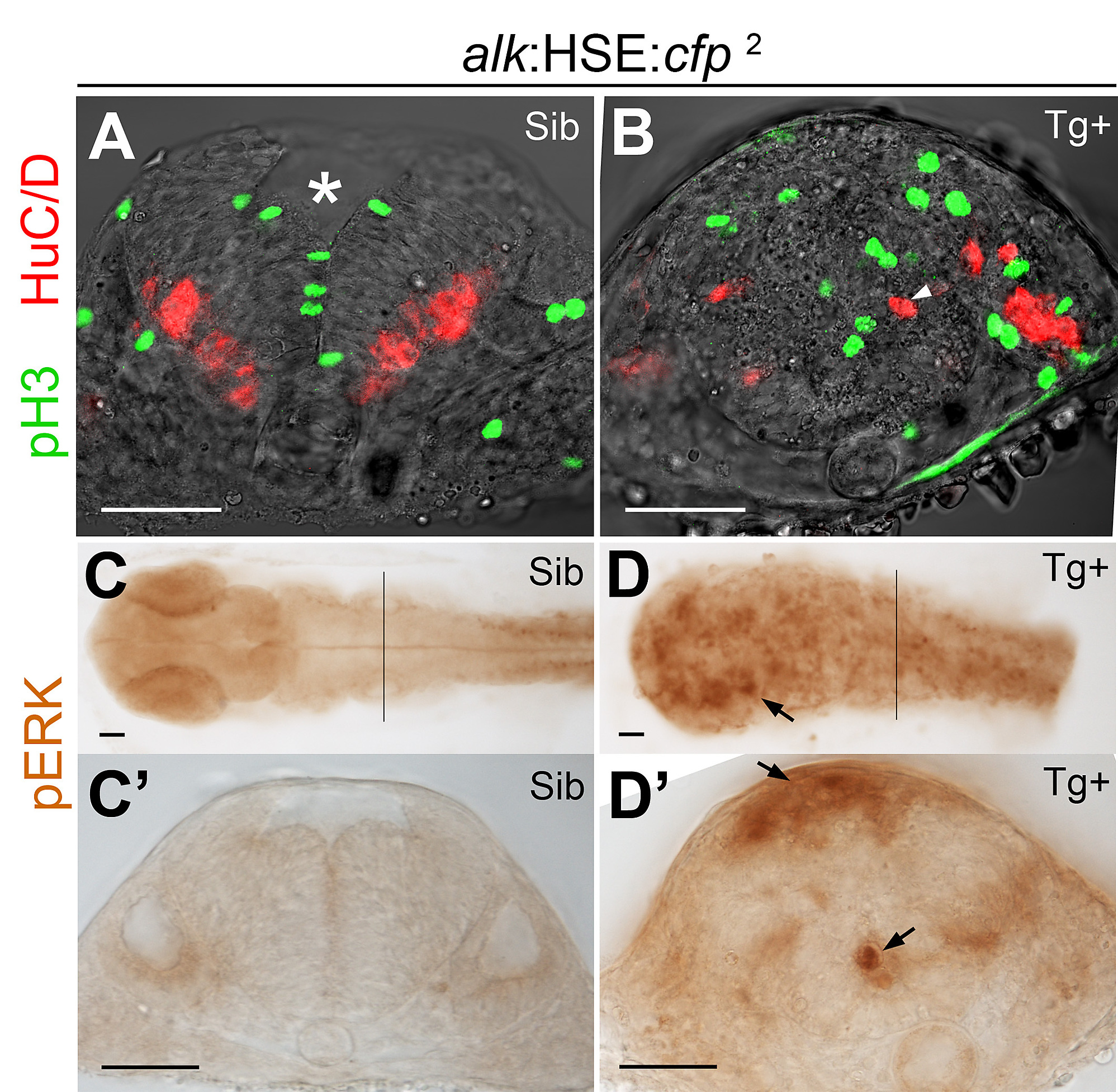
Fig. S4 Proliferation and differentiation in the alk:HSE:cfp2 line. (A,B) Confocal sections of 24hpf embryos of the alk:HSE:cfp2 line. Sib (A) and Tg+ (B) had different neural tube shapes. Dividing cells (pH 3, green) and neurons (HuC/D, red) in Tg+ embryos (B) were both mispositioned (arrowhead), identical to defects observed in the alk:HSE:cfp1 line (Fig. 2). Asterisk labels 4th ventricle. (C,C′,D,D′) Ectopic pERK (arrows) in Tg+ (D,D′) of the alk:HSE:cfp2 line compared to Sib (C,C2), identical to defects observed in alk:HSE:cfp1 (Fig. 3). pERK, phosphorylated-ERK1/2. Sib, transgene negative siblings. Tg+, transgenic embryos. Scale bars: 50 μm.