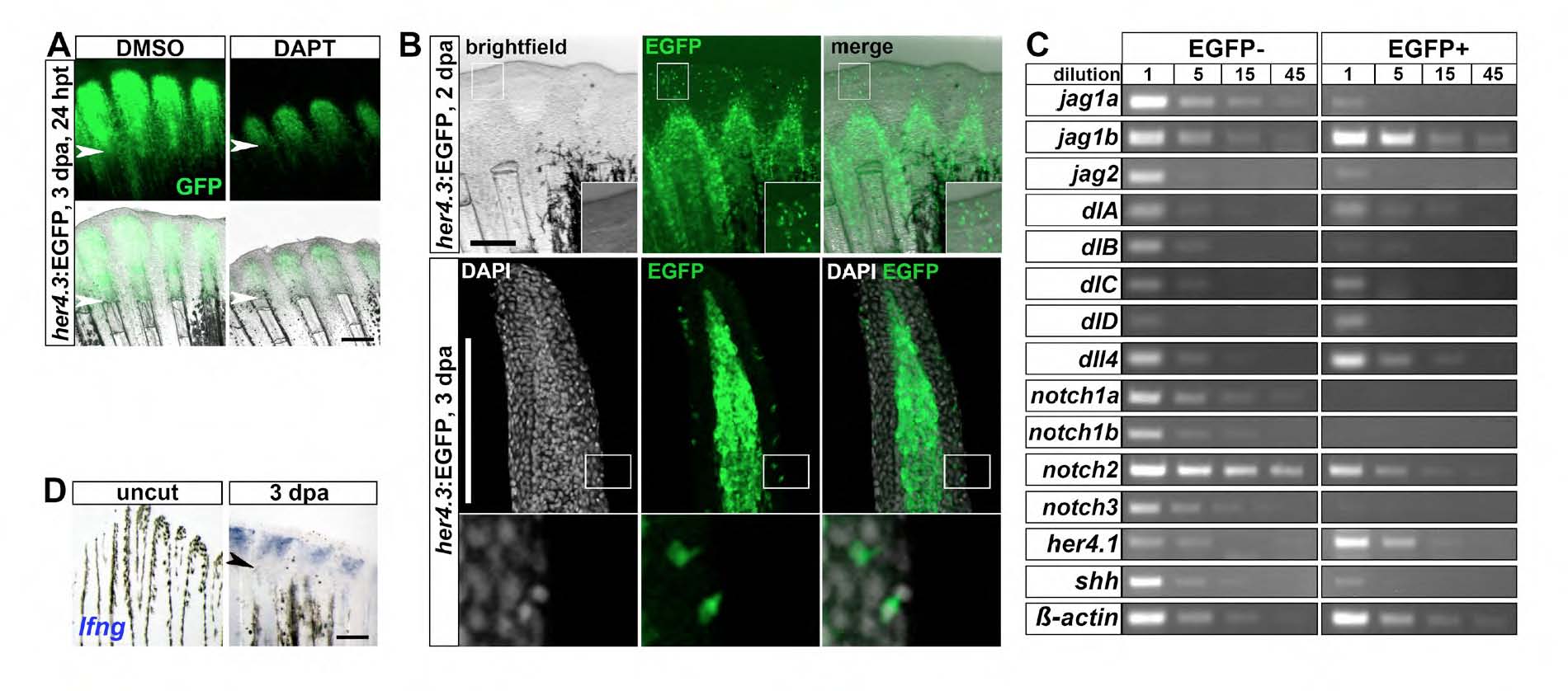
Fig. S1 Expression of a transgenic reporter for Notch signaling and Notch receptors and ligands during fin regeneration. (A) In her4.3:EGFP transgenic regenerates treated with DAPT for 24 hours (24 hpt), EGFP fluorescence is reduced relative to DMSOtreated fins (n=3/3). (B) EGFP fluorescence in her4.3:EGFP transgenic regenerates is detectable in a few scattered cells in the wound epidermis. Top panel: whole-mount image of regenerates at 2 dpa. Bottom panel: confocal images of longitudinal sections of regenerates stained with GFP antibody and DAPI at 3 dpa. (C) Semi-quantitative PCR of the indicated genes on serial dilutions of cDNA derived from the EGFP-positive and EGFP-negative cellular pools of FACS-sorted her4.3:GFP regenerates at 3 dpa. (D) lunatic fringe (lfng) expression, as detected by whole-mount in situ hybridization, is upregulated in the regenerating fin at 3 dpa relative to uncut fins. Arrowheads indicate amputation plane. Scale bars: 200 μm.
Image
Figure Caption
Acknowledgments
This image is the copyrighted work of the attributed author or publisher, and
ZFIN has permission only to display this image to its users.
Additional permissions should be obtained from the applicable author or publisher of the image.
Full text @ Development

