
IMAGE
Fig. 1
- ID
- ZDB-IMAGE-130607-34
- Publication
- Kimura et al., 2013 - Hindbrain V2a Neurons in the Excitation of Spinal Locomotor Circuits during Zebrafish Swimming
- All Figures
- Figures for Kimura et al., 2013
Image
|
Figure Caption
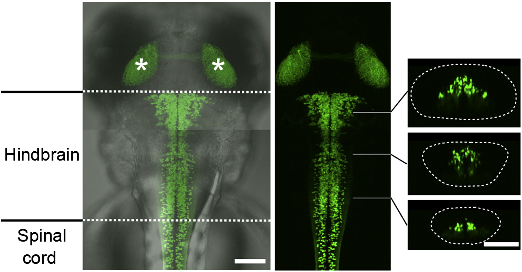
Fig. 1 V2a Neurons in the Hindbrain Compound transgenic fish of Tg[chx10:Gal4] and Tg[UAS:GFP] at 3 dpf. The two panels on the left are dorsal views. GFP-expressing neurons are present in the hindbrain and the spinal cord. The asterisks show the GFP signal in the tectums that are originated from GFP-positive cells in the retina. The three panels on the right are cross-sections. The dotted circle demarcates the hindbrain. Scale bar represents 100 μm.
Acknowledgments
This image is the copyrighted work of the attributed author or publisher, and
ZFIN has permission only to display this image to its users.
Additional permissions should be obtained from the applicable author or publisher of the image.
Full text @ Curr. Biol.