
Fig. S2
- ID
- ZDB-IMAGE-130607-32
- Publication
- Kimura et al., 2013 - Hindbrain V2a Neurons in the Excitation of Spinal Locomotor Circuits during Zebrafish Swimming
- All Figures
- Figures for Kimura et al., 2013
|
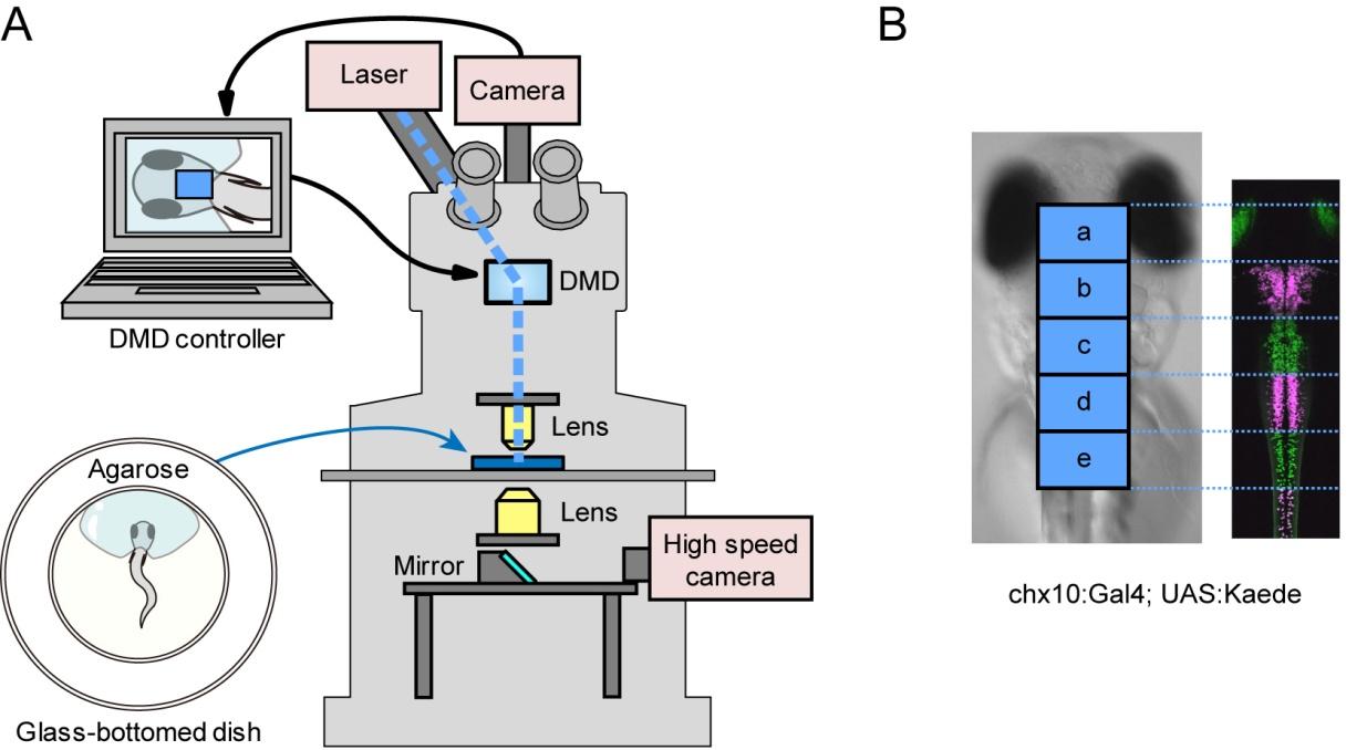
Fig. S2 Photo-stimulation of V2a neurons (A), A schematic of the experimental setup. A larva was mounted on agarose with its tail left free. The region and duration of the photo-stimulation is controlled by digital micro-mirror device (DMD) that is attached to the upright epifluorescent microscope. Movements of the larva are monitored from the bottom using a high-speed camera. (B), Demonstration of region-specific illuminations using Kaede transgenic fish (Tg[chx10:Gal4] and Tg[UAS:Kaede] at 3 dpf). Areas boxed with “a–e” correspond to regions of the photo-stimulations in the ChR experiments. In this larva, regions “b”, “d,” and the spinal cord caudal to region “e” were illuminated with violet light. Magenta represents photo-converted Kaede.