
Fig. 4
- ID
- ZDB-IMAGE-130516-9
- Publication
- O'Neill et al., 2013 - BMP signaling and spadetail regulate exit of muscle precursors from the zebrafish tailbud
- All Figures
- Figures for O'Neill et al., 2013
|
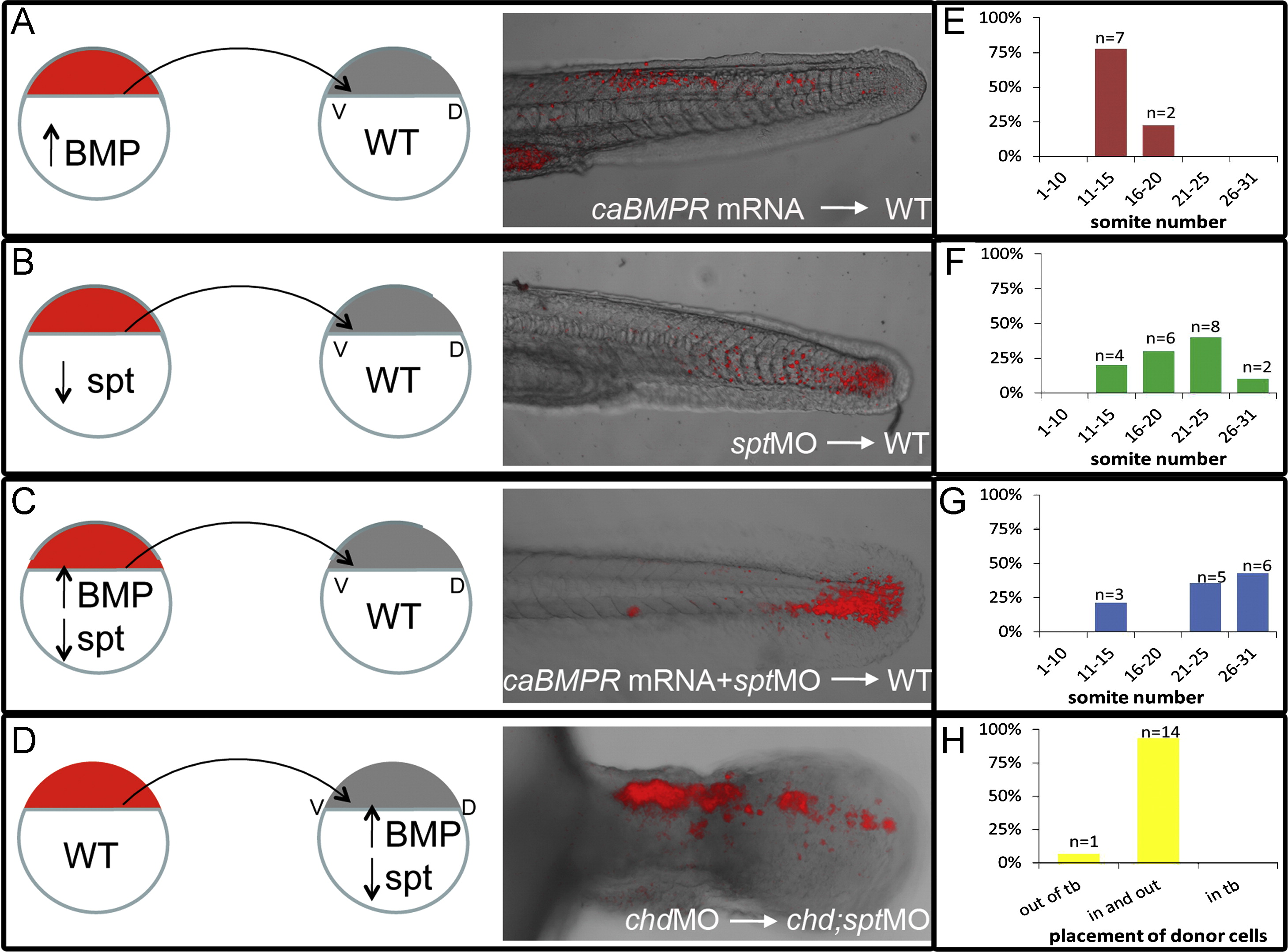
Fig. 4 Ability to exit the tailbud is a cell autonomous fate decision. Cell transplants were performed at 30–50% epiboly stages as diagramed above. Labeled donor cells were placed on the ventral lateral margin of unlabeled host embryos. (A)–(D), Live embryos were photographed using confocal microscopy at 30–48 hpf. (E)–(H), Recipient embryos were scored based on the most anterior somite containing donor cells. Somite 1 being the most anterior and 31 being the most posterior. Cells injected with caBMPR mRNA 4 μg/mL and placed in a WT host were able to exit the tailbud and differentiate in posterior and anterior tail somites, n=9 ((A) and (E)). Cells injected with sptMO 3 μg/mL were also able to leave the tailbud in WT background and contribute to tail somites, n=20 ((B) and (F)). Cells injected with caBMPR mRNA +sptMO were not able to efficiently leave the tailbud in WT backgrounds. A few donor cells could be occasionally found in posterior tail somites and 3 embryos had donor cells in anterior tail somites. However, the majority of transplanted cells were still in the tailbud at 48 hpf, n=14 ((C) and (G)). When chdMO cells were placed in a chdMO;sptMO background they were able to exit the tailbud and migrate anteriorly, n=15 ((D) and (H)).
Reprinted from Developmental Biology, 375(2), O'Neill, K., and Thorpe, C., BMP signaling and spadetail regulate exit of muscle precursors from the zebrafish tailbud, 117-127, Copyright (2013) with permission from Elsevier. Full text @ Dev. Biol.