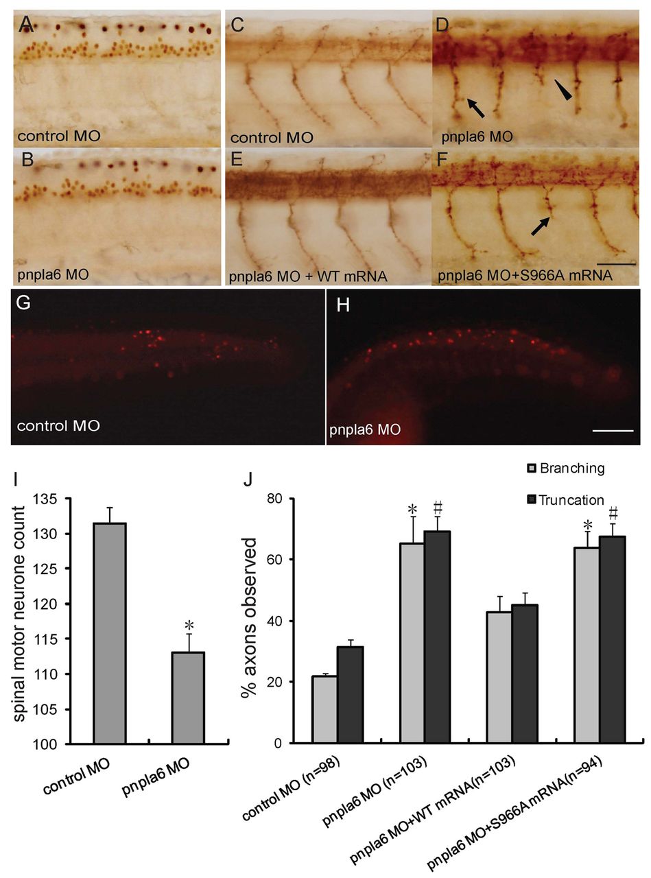
Fig. 3 Knockdown of pnpla6 leads to primary motor neuron defect. (A,B) Lateral views of whole-mount embryos labeled with islet1 mAb. Zebrafish pnpla6 morphants (B) showed fewer motor neurons than control animals (A). (C-F) Lateral views of whole-mount embryos labeled with mAb Znp-1: 36-hpf embryo injected with control MO (C); 36-hpf pnpla6 MO morphant (D) shows truncated (arrow) motor axons, and branching of (arrowhead) motor axons. The defects including truncation and branching can be rescued by human wild-type mRNA (E) but not by mutant mRNA (F). (G,H) Apoptosis analysis of pnpla6 morphants. Zebrafish pnpla6 morphant (H) and control (G) embryos were analyzed by TUNEL assay. (I) Quantification of motor neurons in pnpla6 morphants and controls. The total number of islet1-positive nuclei in the spinal cord spanning ten somites on one side of the spinal cord was counted. Values given are means + s.e.m. and 6–8 individual embryos were included in each group; *P<0.05 compared with control. (J) Quantification of the motor axon defects (truncation or branching) in four groups of embryos: pnpla6 MO, control MO, pnpla6 MO + human wild-type PNPLA6 mRNA and pnpla6 MO + human mutant PNPLA6 mRNA at 26 hpf. #P<0.05 compared with control. Scale bars: 50 μm (A-F); 200 μm (G,H).
Image
Figure Caption
Figure Data
Acknowledgments
This image is the copyrighted work of the attributed author or publisher, and
ZFIN has permission only to display this image to its users.
Additional permissions should be obtained from the applicable author or publisher of the image.
Full text @ Dis. Model. Mech.

