
Fig. S3
- ID
- ZDB-IMAGE-130516-21
- Publication
- Schuler et al., 2013 - Ciliated sensory hair cell formation and function require the F-BAR protein syndapin I and the WH2 domain-based actin nucleator Cobl
- All Figures
- Figures for Schuler et al., 2013
|
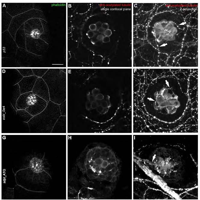
Fig. S3 Morphology and innervations of mature neuromasts (5 dpf) are similar in controls and in cobl and syndapin I morphant larvae. (A,D,G) Phalloidin staining of mature neuromasts (5 dpf) visualizing the overall organization of neuromast rosettes of the posterior lateral line organ. Controls (A) as well as cobl (D) and syndapin I morphants (G) display properly polarized neuromasts. Single confocal planes illustrate the regular morphology of the cell clusters. (B,E,H,C,F,I) Immunolabeling with antiacetylated tubulin stains cell bodies, kinocilia and axons. Control, cobl- and syndapin I-morphant neuromasts are contacted by axons, as can be seen in the corresponding Z-projections of confocal images (C,F,I; marked by arrows). Bar, 10 μm.