
Image
|
Figure Caption
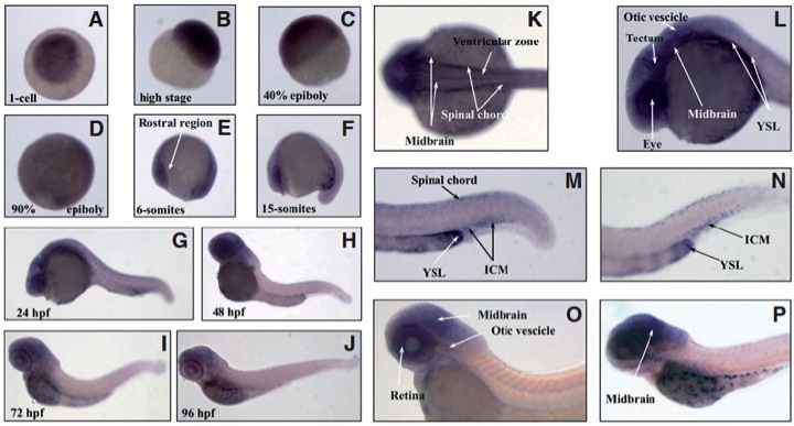
Fig. 10 (A-J) Whole-mount in situ hybridization of mcoln1.1 at different stages of zebrafish development (63X magnification). Dorsal (K) and lateral (L) view of 24 hpf embryo head. Magnification of 24 hpf (M) and 36 hpf (N) embryo tails. Lateral view of 48 hpf (O) and 80 hpf (P) embryo. Images (K-P) were acquired at 115X magnification. Relevant sites of mcoln1.1 gene expression are indicated with arrows.
Figure Data
Acknowledgments
This image is the copyrighted work of the attributed author or publisher, and
ZFIN has permission only to display this image to its users.
Additional permissions should be obtained from the applicable author or publisher of the image.
Full text @ Int. J. Dev. Biol.