
Fig. 3
- ID
- ZDB-IMAGE-130509-24
- Publication
- Yang et al., 2013 - A novel zebrafish xenotransplantation model for study of glioma stem cell invasion
- All Figures
- Figures for Yang et al., 2013
|
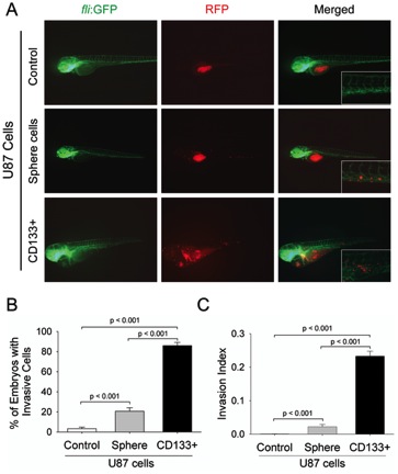
Fig. 3 CD133+ U87 GSCs are highly invasive within zebrafish embryos.
A. Representative images of the invasion of differentiated U87 cells, U87 sphere cells, and CD133+ U87 GSCs within the injected embryos at 2 dpi. The images at higher magnification show the invasive RFP-labeled cell masses at tail region of embryos via host vessels. B. The percentage of the embryos with invasive cells injected with RFP-labeled differentiated U87 cells, U87 sphere cells, and CD133+ U87 GSCs. The data were obtained from three replicate experiments of 50 injected embryos in each experiment: n = 124 for live embryos injected with differentiated U87 cells, n = 121 for embryos injected with U87 sphere cells, and n = 120 for embryos injected with CD133+ cells C. The percentage of invasive cells within total injected cells (Invasion Index) in the embryos. All injected cells including invasive or non-invasive cells within zebrafish embryos were evaluated by ImageJ software through fluorescence intensity. n = 37200 (300 injected cells per embryo among 124 live embryos) for differentiated U87 cell group, n = 36300 (300 injected cells per embryo among 121 live embryos) for U87 sphere cell group, and n = 36000 (300 cells per embryo among 120 live embryos) for CD133+ U87 GSCs group (p<0.001).