
Fig. 2
- ID
- ZDB-IMAGE-130509-23
- Publication
- Yang et al., 2013 - A novel zebrafish xenotransplantation model for study of glioma stem cell invasion
- All Figures
- Figures for Yang et al., 2013
|
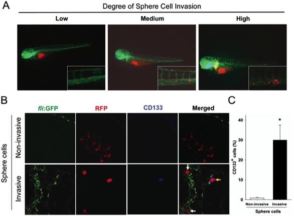
Fig. 2 Invasive U87 sphere cells express CD133.
A. U87 sphere cells with various invasion capability within zebrafish embryos. The extent of invasion was classified in three degrees: Low: less than 5 migrated cells; Medium: 5–20 migrated cells; High: more than 20 migrated cells. Representative images at higher magnification show the invasive RFP-labeled U87 sphere cell masses (red) in the tail region of the embryos via EGFP-labeled host vessels (green). B. Detection of CD133 expression on non-invasive and invasive U87 sphere cells at 2 dpi by immunofluorecent staining. All of U87 sphere cells within injected embryos were stained with monoclonal anti-CD133 antibody (1:300) and examined by confocal microscopy. Green: Tg (fli1:EGFP)y1 microvessels; red: RFP-labeled U87 sphere cells; blue: CD133 positive U87 cells. C. Quantitative analysis of CD133-expressing cells in non-invasive cell group (n = 713) and high-invasive cell group (n = 175) at 2 dpi. (p<0.001).