
Fig. 1
- ID
- ZDB-IMAGE-130509-22
- Publication
- Yang et al., 2013 - A novel zebrafish xenotransplantation model for study of glioma stem cell invasion
- All Figures
- Figures for Yang et al., 2013
|
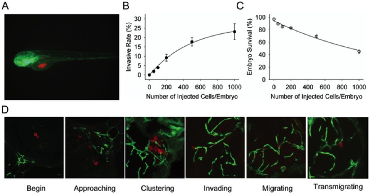
Fig. 1 The establishment of U87 glioma sphere cell invasion model in zebrafish embryos.
A. Dual color confocal image shows that U87 sphere cells (RFP labeled, red) were microinjected into the middle of yolk sac within Tg (fli1:EGFP)y1 transgenic zebrafish embryos (EGFP labeled, green). B. Different numbers of U87-RFP glioma sphere cells were microinjected into Tg (fli1:EGFP)y1 embryos (n = 300 in each group), and the percentage of embryos with invasive tumor cells was quantitated. C. The survival rate of Tg (fli1:EGFP)y1 zebrafish embryos microinjected with different numbers of U87-RFP glioma sphere cells (n = 300 in each group). D. Representative dual color confocal images of RFP-labeled U87 sphere cells within Tg (fli1:EGFP)y1 zebrafish embryos at the different invasive stages. Red: RFP-labeled U87 sphere cells; Green: Tg (fli1:EGFP)y1 microvessels.