
Fig. S2
- ID
- ZDB-IMAGE-130507-12
- Publication
- Basten et al., 2013 - Mutations in LRRC50 Predispose Zebrafish and Humans to Seminomas
- All Figures
- Figures for Basten et al., 2013
|
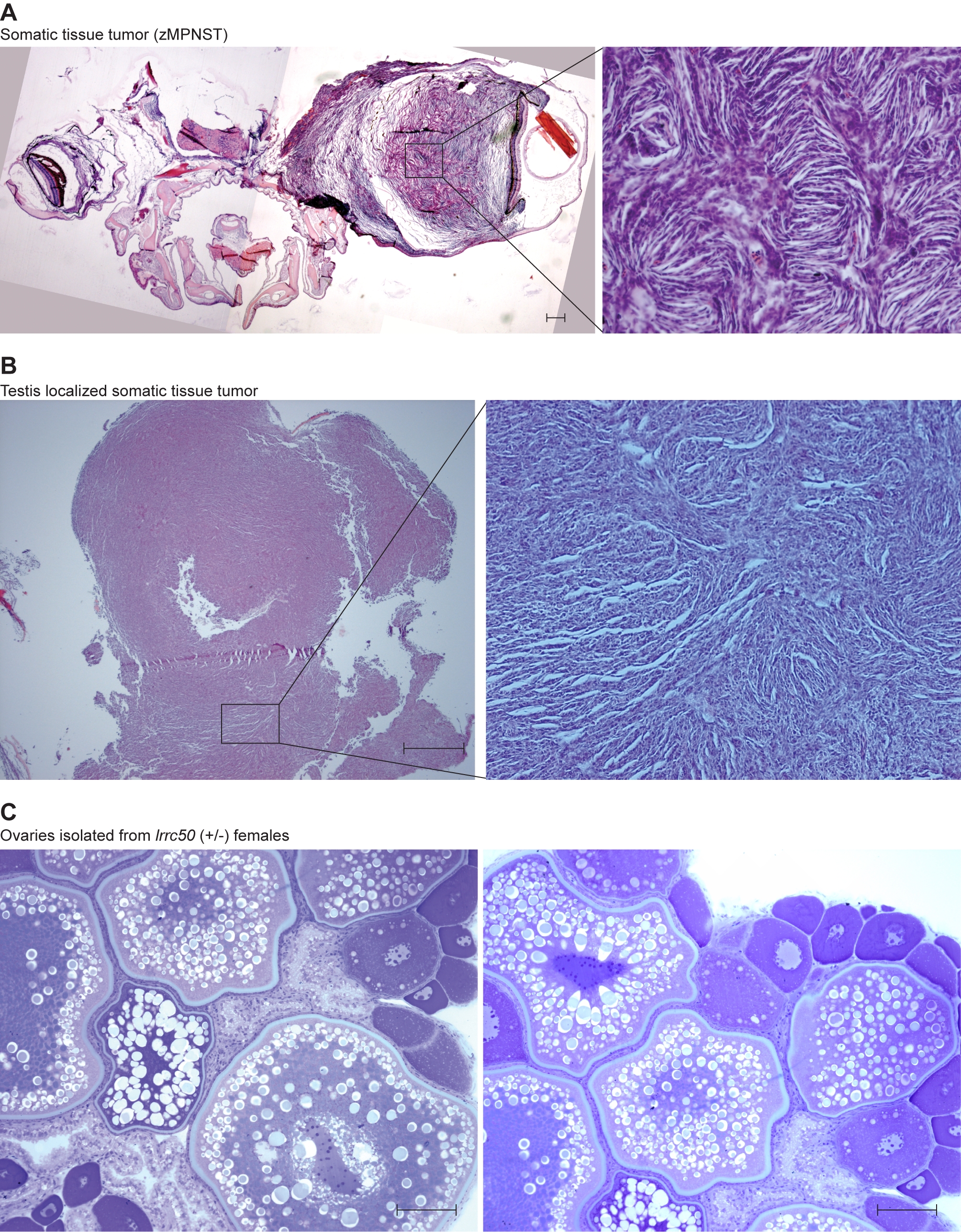
Fig. S2
Non-TGCT zebrafish lrrc50Hu255h tumors and female gonad. (A) One fish was identified bearing a large tumor (merge of two images) located proximal to the brain, histologically resembling zebrafish malignant peripheral nerve sheath tumors (zMPNST). Albeit a rare finding in lrrc50Hu255h zebrafish, these tumors typically do not occur in wild-type zebrafish and might therefore potentially represent an alternative lrrc50 associated tumor type. Scale bars; 50 μm. (B) We identified a tumor of somatic tissue (undetermined pathology, putative zMPNST) located in the testes of one zebrafish, unlike all other TGCTs described in this manuscript. Scale bars; 50 μm. (C) Heterozygote lrrc50 females (n = 11) do not show gonadal abnormalities; ovaries were isolated simultaneously with male testes/TGCT at an age of 30 months. Scale bars; 50 μm.