Fig. 6
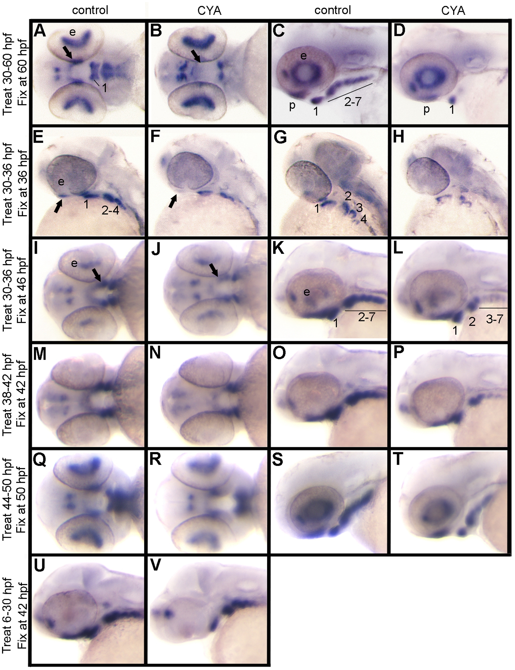
Continuous Hh signaling is required for satb2 expression.
Ventral (A, B, I, J, M, N, Q, R), lateral (C, D, E, F, K, L, O, P, S, T, U, V) and dorsal-lateral (G, H) images of satb2 expression by in situ hybridization are shown in control and cyclopmaine-treated (CYA) embryos. (A, C) In controls at 60 hpf, satb2 is expressed in palatal precursors and in the ventral region of each arch. (B, D) Embryos treated with cyclopamine from 30–60 hpf show a dramatic reduction of satb2 expression in the palatal precursors, pharyngeal arch 1 and 2 as well as complete loss of expression in the posterior arches. (E–H) Compared to controls, embryos treated with cyclopamine from 30 to 36 hpf also show reduction of satb2 expression in the palatal precursors and arches 1 and 2, with a more complete loss of expression in the more posterior arches. (I–L) If, however, embryos are removed from cyclopamine at 36 hpf and allowed to develop for 10 hours, there is a partial recovery of satb2 expression. (M–T) Cyclopamine treatment either from 38 to 42 hpf (M–P) or 44 to 50 hpf (Q–T) results in mild reduction of satb2 expression in the palatal precursors and pharyngeal arches. (U, V) While the maxillary domain is lost in embryos treated with cyclopamine from 6–30 hpf the expression in the ventral arches appears largely intact. CYA, cyclopamine; e, eye; b, brain; p, palatal precursors; pharyngeal arches are numbered in some panels for clarity.

