
Fig. S2
- ID
- ZDB-IMAGE-130502-10
- Publication
- Song et al., 2013 - Pou5f1-dependent EGF expression controls e-cadherin endocytosis, cell adhesion, and zebrafish epiboly movements
- All Figures
- Figures for Song et al., 2013
|
Fig. S2
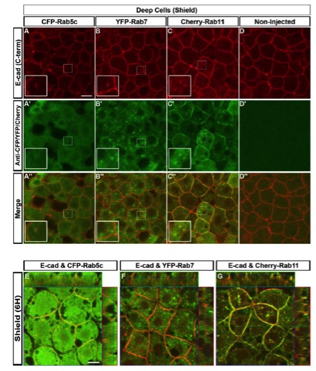
E-cad Immunoreactive Endosomes Colocalize with Rab5c, and 11, but not with Rab7 (Related to Figure 2)
(A-D′ ′) Double immunofluorescence detection of C-term E-cad (A-D, red) and CFP-Rab5c, YFP-Rab7, and Cherry-Rab11 fusion proteins (A′-D′, green) in deep cells of shield stage WT embryos. (A′ ′-D′ ′) Merged channels reveal colocalization of E-cad to Rab5c and Rab11 immunoreactive endosomes, but not to Rab7 endosomes. Insets show higher magnification views of the boxes. Animal views. Scale bar = 10 μm.
(E-G) 3D reconstruction of confocal image stacks showing double immunofluorescence for E-cad and 6 CFP-Rab5c, YFP-Rab7, or Cherry-Rab11. E-cad co-localizes with CFP-Rab5c (E) or Cherry-Rab11 (G) immunoreactive endosomes both at the plasma membrane and intracellular vesicles, but not to YFP-Rab7 immunoreactive endosomes (F). Animal views. Scale bar = 10 μm.
Reprinted from Developmental Cell, 24(5), Song, S., Eckerle, S., Onichtchouk, D., Marrs, J.A., Nitschke, R., and Driever, W., Pou5f1-dependent EGF expression controls e-cadherin endocytosis, cell adhesion, and zebrafish epiboly movements, 486-501, Copyright (2013) with permission from Elsevier. Full text @ Dev. Cell