
Fig. S6
- ID
- ZDB-IMAGE-130429-11
- Publication
- Liu et al., 2013 - Direct and indirect roles of Fgf3 and Fgf10 in innervation and vascularisation of the vertebrate hypothalamic neurohypophysis
- All Figures
- Figures for Liu et al., 2013
|
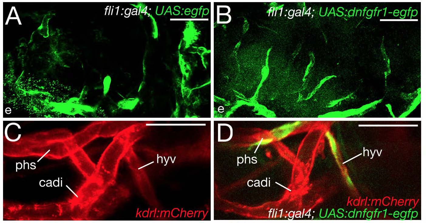
Fig. S6 Endothelial cells expressing the dominant-negative Fgfr1 receptor contribute normally to other cephalic blood vessels and do not affect morphogenesis of the hypophyseal vein. (A-D) Confocal in vivo images of different transgenic fish, as indicated; 100 hpf; ventral views, anterior to the left. Injection of the UAS:egfp control (A) or the UAS:dnfgfr1-egfp plasmid (B,D) into tg(fli1a:gal4); tg(kdrl:mcherry) double transgenic animals leads to mosaic expression of EGFP (A) or the truncated Fgfr1 receptor fused to EGFP in endothelial cells. For simplicity, the red channel is omitted in A and B, and the green channel omitted in C. (A,B) Endothelial cells with activation of the UAS-egfp transgene (A) or the UAS:dnfgfr1-egfp transgene (B) display comparable contributions to cephalic blood vessels. Nevertheless, their contributions to the hypophyseal artery and the two branches encompassing the pituitary differ (80% versus 40%; data not shown). (C,D) Morphogenesis of the hypophyseal vein is unaffected by endothelial cells expressing the UAS:dnfgfr1-egfp transgene. cadi, caudal division of internal carotid artery; e, eye; hyv, hypophyseal vein; phs, primary head sinus. Scale bars: 50 μm.