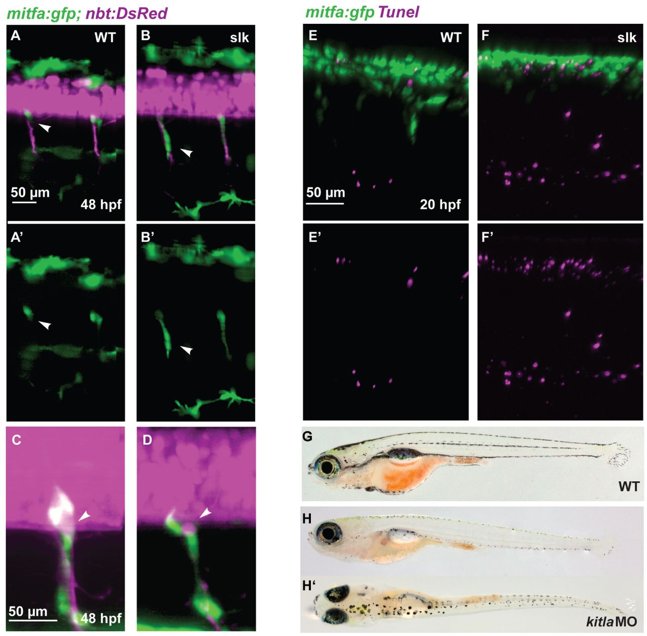
Fig. 9 Abnormal NC migration and apoptosis in slk embryos. (A-B′) GFP-positive cells in double transgenic Tg(mitfa:gfp; nbt:DsRed) wild-type (A,A′) and slk mutant (B,B′) embryos at 48 hpf. In the wild-type embryo, GFP-positive NC cells remain at the position of the DRG and have a rounded morphology (white arrowhead in A,A2) whereas in slk mutants they are stretched along the nerves and appear to be migratory (white arrowheads in B,B2). A′, B′ show green channel only. (C,D) Magnification showing mitfa:gfp-labelled cells at the exit point of the spinal nerves at 48 hpf of wild-type (C) and slk mutant (D) embryos. The arrowheads point to DRGs. (E-F′) TUNEL staining in Tg(mitfa:gfp) embryos in wild-type (E,E′) and slk (F,F′) embryos at 20 hpf. E′,F′ show red channel only. (G-H′) Wild type (G) and slk morphant (H,H′) at 20 dpf (5.7 mm SL).
Image
Figure Caption
Figure Data
Acknowledgments
This image is the copyrighted work of the attributed author or publisher, and
ZFIN has permission only to display this image to its users.
Additional permissions should be obtained from the applicable author or publisher of the image.
Full text @ Development

