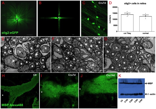
Fig. 5 Oligodendrocytes in retina did not decrease after optic nerve injury.
(A) Scan the whole retina with 10× objective of LSM710. (B) Cruciated scan in 3D way for counting the destiny of olig2+ cells. (C) The processes of oligodendrocytes keep integrality after ONI. (D) Oligodendrocytes number did not decrease at one week after ONT. (E-G) TEM images also show that myelin structure within NFL keeps integrity in ONC or ONT model within the first week. (H-J) Both IHC and western blot results (K) indicated that MBP in retina was not decreased after optic nerve injury, * indicates lens; Scale bar: 200 μm (A); 50 μm (B, H-J), 10 μm (C); 200 nm (E-G).

