
|
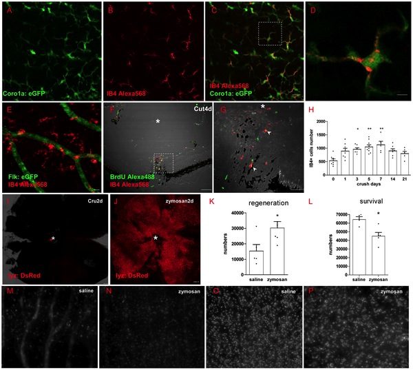
Fig. 3 Inflammation has dual-roles during regeneration
(A-D) IB4+ cells in retina are colocalized with coro1a:eGFP in Tg(coro1a:eGFP; lyz:DsRed) fish, which means IB4+ cells mark microglia cells in zebrafish retina. (D) Shows the detailed view of square in (C). (E) IB4+ cells could not colocalize with flk:GFP in transgenic Tg(flk:GFP) line. (F-G) BrdU marker shows that some IB4+ cells are newborn in retina (arrowhead in G), * indicates the lens. (H) IB4+ cells number in all layers of retina changed with time, increasing significantly 3 days to 7 days after ONC, then decreasing to normal level after 14 days. (I-J) Zymosan induces acute neutrophil/macrophage infiltration into retina, * indicates optic disc. (K, M and N) Neutrophil/macrophage infiltration into retina could increase the number of regenerated RGCs in the first week after ONT, but decrease RGCs survival after 2 weeks (L, O and P). * indicates p<0.05, ** is p<0.01. Scale bar: 10 μm (A-C, M-P); 2 μm (D); 30 μm (G), 100 μm (F, I and J).