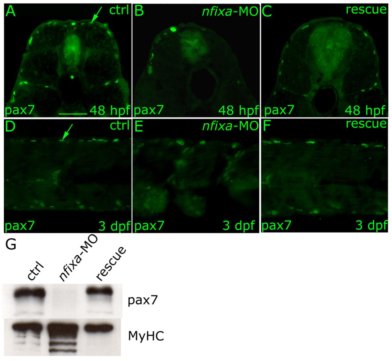
Fig. 7 The absence of Pax7-positive progenitor cells in nfixa-MO-injected embryos determines the loss of secondary fibers. (A-F) Immunohistochemistry experiments with the Pax7 antibody that recognizes secondary fiber precursors. Pax7-positive secondary fiber progenitors at the dorsal and ventral extremes of the superficial muscle monolayer did not form in 48 hpf and 3 dpf nfixa-MO-injected embryos (B,E) in comparison with controls (A,D arrow), whereas they are recovered in rescued embryos (C,F). (G) Western blot: secondary fiber precursors were recognized by Pax7 antibody, all sarcomeric myosins MyHC were recognized by MF20 antibody. In isolated tails of nfixa-MO-injected larvae, Pax7 protein levels were higher than the control whereas they were recovered in rescued embryos at 3 dpf. All myosin protein levels were higher in nfixa-MO-injected larvae, as expected. (A-C) 48 hpf transversal sections, dorsal on top. (D-F) 3 dpf longitudinal sections, anterior towards the left. Scale bar: 100 μm.
Image
Figure Caption
Figure Data
Acknowledgments
This image is the copyrighted work of the attributed author or publisher, and
ZFIN has permission only to display this image to its users.
Additional permissions should be obtained from the applicable author or publisher of the image.
Full text @ Development

