
Image
|
Figure Caption
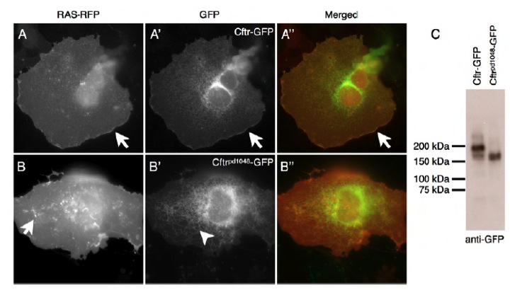
Fig. S1 Cftrpd1048-GFP is mislocalized. (A-A′′) Zebrafish Cftr-GFP expressed in Cos-7 cells is predominantly localized to the plasma membrane. (B-B′′) The Cftrpd1048-GFP fusion protein has a reduction in plasma membrane localization. Arrows point to membrane localization, arrowheads denote the internal pool. (C) A western blot for GFP detects Cftr-GFP and Cftrpd1048-GFP expressed from HEK293 cells. Scale bars: 50 μm.
Acknowledgments
This image is the copyrighted work of the attributed author or publisher, and
ZFIN has permission only to display this image to its users.
Additional permissions should be obtained from the applicable author or publisher of the image.
Full text @ Development