
Fig. 4
- ID
- ZDB-IMAGE-130416-4
- Publication
- Schmidt et al., 2013 - Early stages of retinal development depend on Sec13 function
- All Figures
- Figures for Schmidt et al., 2013
|
Fig. 4
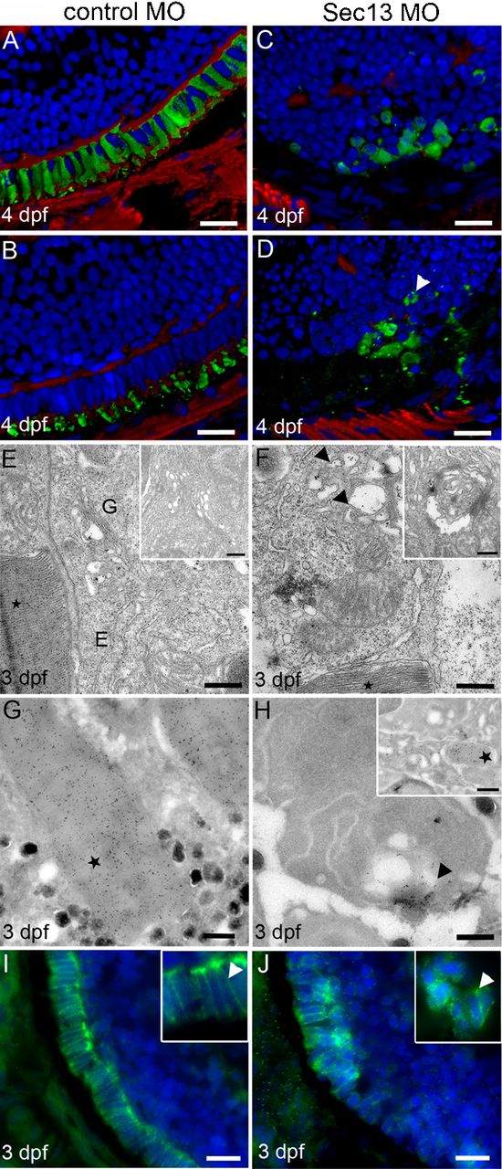
Photoreceptor development and opsin transport in Sec13 morphant eyes.
3D opacity view of control (A,B) and Sec13 MO (C,D) stained for cones (zpr-1, A,C) and opsin (B,D) (green), phalloidin (red) and DAPI (blue). Arrowhead: dotted opsin localisation in Sec13 MO. Scale bars: 10μm. TEM of control (E) and Sec13 MO (F), see insets for additional examples. Stars: outer segments; E: ER; G: Golgi; arrowheads: dilated ER and Golgi. Scale bars: 500nm. Immunogold-labelling of opsin. Control (G) and Sec13MO (H). Arrowhead: accumulation of opsin in cell periphery. Stars: opsin label in outer segments. Scale bars: 500nm. Immunolocalisation of syntaxin-3 in control (I) and Sec13 MO (J). Arrowheads: syntaxin-3 label at plasma membrane. Scale bars: 10μm.