
Fig. 8
- ID
- ZDB-IMAGE-130416-18
- Publication
- Navis et al., 2013 - Cftr controls lumen expansion and function of Kupffer's vesicle in zebrafish
- All Figures
- Figures for Navis et al., 2013
|
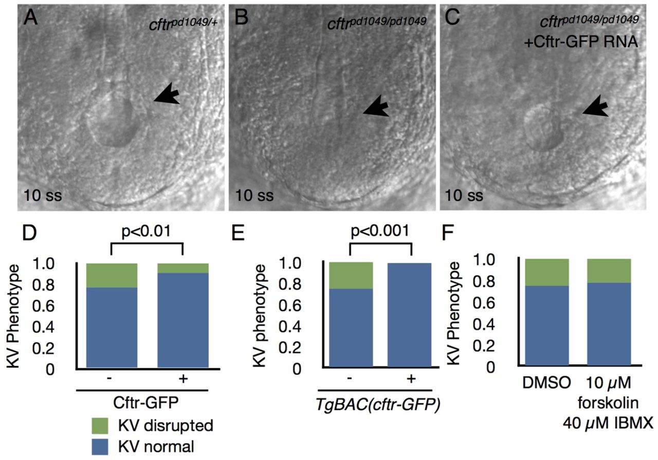
Fig. 8 Expression of Cftr-GFP can rescue lumen expansion defects in cftr mutants. (A-C) Representative DIC images of KV (arrows) at 10 ss in embryos (A) heterozygous for cftrpd1049, (B) homozygous for cftrpd1049 and (C) in cftrpd1049 homozygous mutants injected with 150 pg cftr-GFP RNA. (D) Quantification of KV phenotype at 10 ss in control and cftr-GFP-injected embryos resulting from a cross between cftrpd1049 heterozygous parents. Control, n=158; Cftr-GFP, n=94; P<0.01. (E) Graph of the KV phenotype at 10 ss in embryos from a cftrpd1049/+; TgBAC(cftr-GFP) × cftrpd1049/+ cross, compared by whether the embryos were Cftr-GFP positive or negative. Cftr-GFP negative, n=140; Cftr-GFP positive, n=149; P<0.001. (F) Quantification of KV phenotype in embryos treated with DMSO (n=96) or 10 μM forskolin and 40 7mu;M IBMX resulting from a cross between cftrpd1049 heterozygous parents. n=95; P=0.6374. Scale bars: 50 μm.