
|
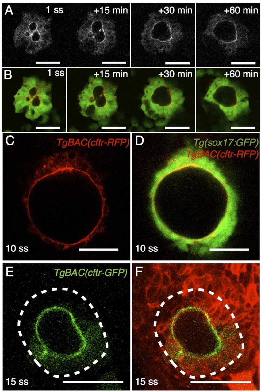
Fig. 6 Cftr is apically localized in KV epithelial cells throughout its morphogenesis. (A) Live, time-lapse confocal imaging of Cftr-RFP in TgBAC(cftr-RFP); Tg(sox17:GFP) embryos. Cftr-RFP is expressed and apically localized in KV throughout the initial stages of lumen coalescence. (B) Merge of the RFP and GFP channels. (C,D) Live confocal imaging of TgBAC(cftr-RFP); Tg(sox17:GFP) embryos at 10 ss shows that Cftr-RFP is localized apically in KV. (D) Merged view of Cftr-RFP and GFP. (E,F) Live confocal imaging of TgBAC(cftr-GFP) embryos injected with membrane-RFP RNA shows continued apical localization of Cftr-GFP until 15 ss. The dashed line marks the edge of KV. Scale bars: 50 μm.