
|
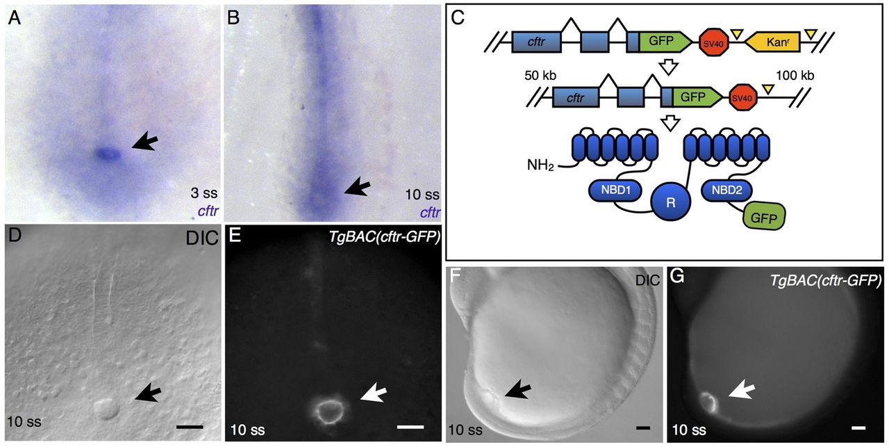
Fig. 5 cftr expression is enriched in KV. (A) At 3 ss, in situ hybridization detects cftr expression specifically in KV (arrow). (B) In situ hybridization showing cftr expression in KV (arrow) and the chordamesoderm in 10 ss embryos. (C) Schematic of the BAC recombineering procedure, showing the recombination target and the expected structure of the resulting GFP fusion protein. (D,F) DIC images showing ventral (D) and lateral (F) views of 10 ss embryos expressing TgBAC(cftr-GFP). The arrow marks the characteristic KV structure. (E,G) Whole-mount epifluorescence of the embryos shown in D and F demonstrate specific KV expression of Cftr-GFP (arrows) at 10 ss. Scale bars: 50 μm.