
|
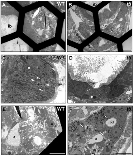
Fig. S6 Absence of dead cells in the intestinal lumen of WT and ttis450 larvae at 7 dpf. (A–F) Transmission electron micrographs of transverse sections of WT and ttis450 larvae at 168 hpf (7 dpf). The number of conspicuous autophagosome-like structures in the IECs of ttis450 larvae has diminished by 7 dpf and there are no dead cells in the lumen (D). Meanwhile, liver cells of ttis450 larvae contain abundant autolysosome-like structures at this time-point (F, white arrows). Scale bars = 50 μm (A, B); 10 µm (C–F). ib, intestinal bulb; n, nucleus; m, mitochondria; mv, microvilli; l, liver; bd, bile duct; a, arteriole.