
|
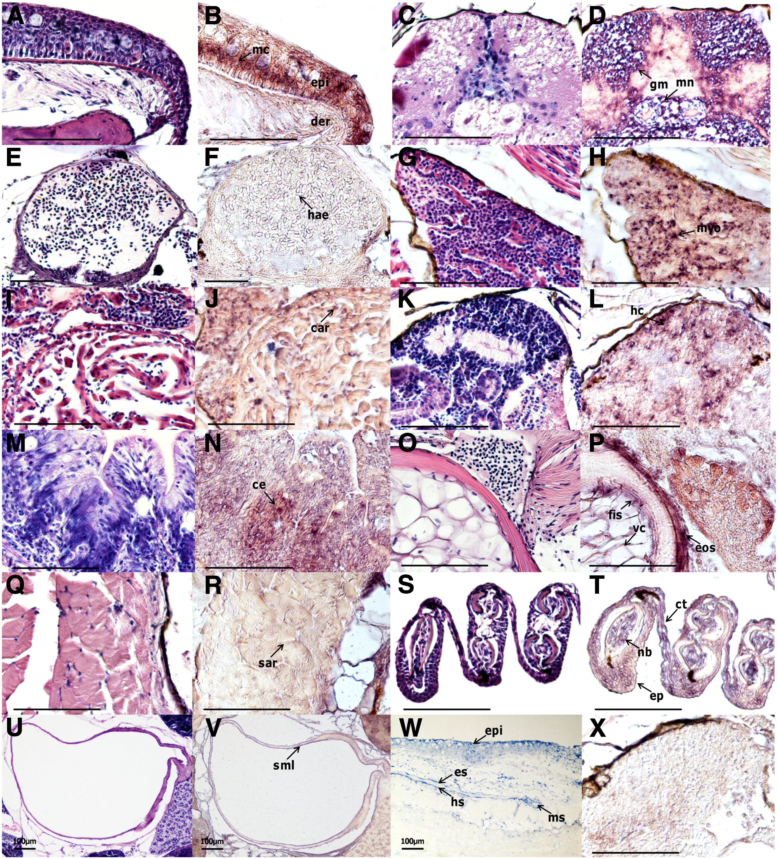
Fig. 5 In situ hybridization of DPT mRNA in zebrafish tissues. Hematoxylin and eosin staining in adult zebrafish tissues: (A) skin; (C) spinal cord; (E) dorsal aorta; (G) atrium; (I) ventricle; (K) kidney; (M) gut; (O) notochord; (Q) muscle; (S) fin; (U) bladder. (B, D, F, H, J, L, N, P, R, T, and V) DPT mRNA detection in zebrafish tissues as a counterpart to A, C, E, G, I, K, M, O, Q, S, and U. (W) In situ hybridization of DPT in the regenerating scale; 7 days after scale removal. (X) Kidney section was hybridized with sense DPT probe as a negative control. Abbreviations: mc, Malpighian cell; der, dermis; epi, epidermis; mn, Mauthner neurons; gm, gray matter; hae, hematocyte; myo, myocardium; car, cardiomyocyte; hc, hematopoietic cell; ce, columnar epithelium; vc, vacuolated cell; fis, fibrous inner sheath; eos, elastic outer sheath; sar, sarcolemma; ct, connective tissue; nb, nerve bundle; ep, epithelium; sml, smooth muscle layer; es, episquamal scleroblast; hs, hyposquamal scleroblast; ms, marginal scleroblast. Scale bar = 100 μm.
Reprinted from Gene, 516(2), Tan, Y., Iimura, K., Sato, T., Ura, K., and Takagi, Y., Spatiotemporal expression of the dermatopontin gene in zebrafish Danio rerio, 277-284, Copyright (2013) with permission from Elsevier. Full text @ Gene