
|
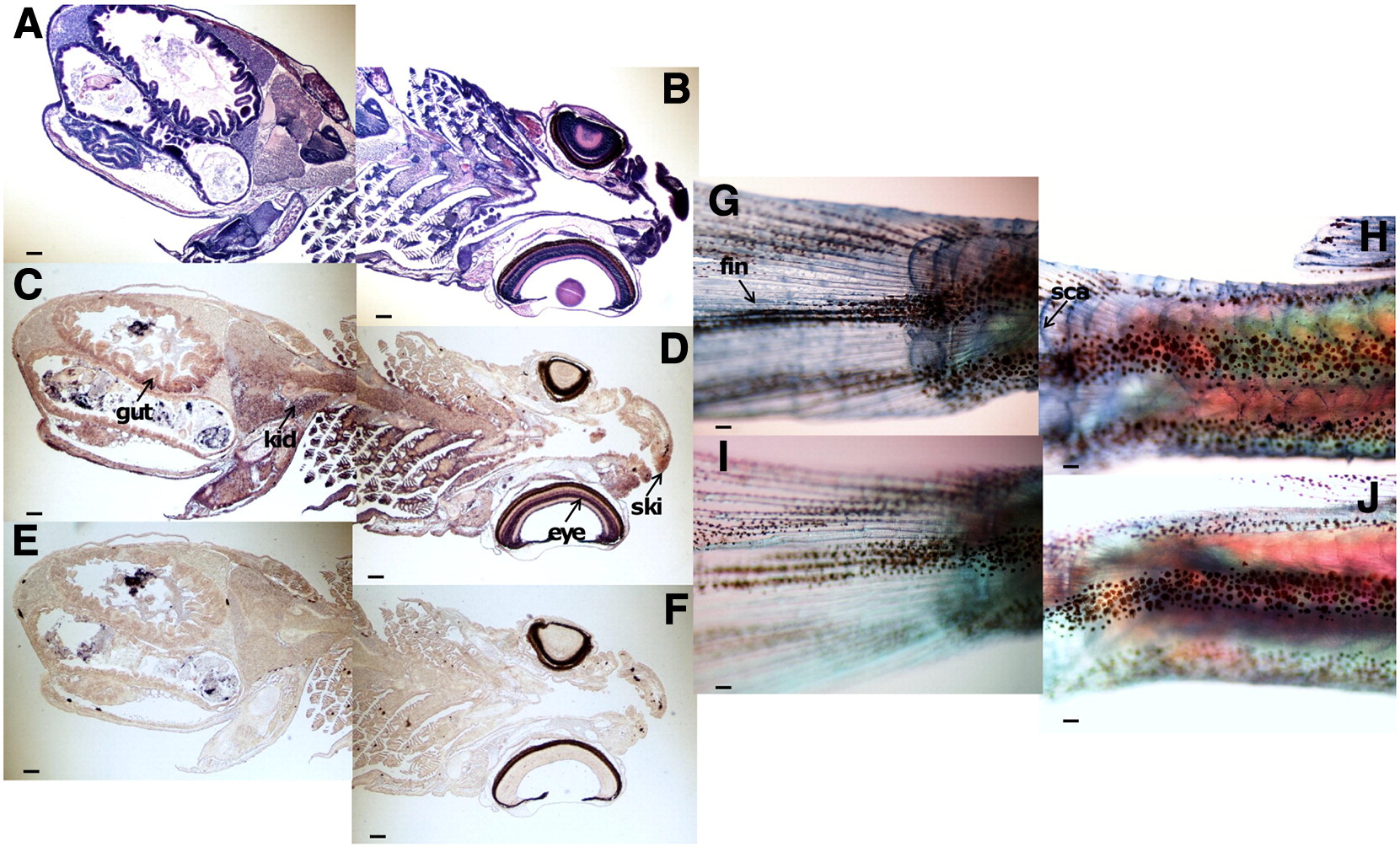
Fig. 6 In situ hybridization of dermatopontin (DPT) mRNA in zebrafish larvae 28 dpf. In these figures, sections were cut frontally from rostral to caudal. The sections (8 µm) were hybridized with the DPT probes. Scale bar is shown bottom left. (A, B) Representative sections of larvae 28 dpf stained using hematoxylin and eosin staining. (C, D) In situ hybridization with DPT antisense probe. DPT mRNA expression was observed in the gut, kidney (C), eye, and skin (D). (E, F) In situ hybridization with DPT sense probes. (G, H) Whole-mount in situ hybridization of the tail zone with DPT antisense probe. (I, J) Whole-mount in situ hybridization of the tail zone with DPT sense probe. Abbreviations: gut, gut; kid, kidney; ski, skin; eye, eye; fin, fin; sca, scale; scale bar = 100 μm.
Reprinted from Gene, 516(2), Tan, Y., Iimura, K., Sato, T., Ura, K., and Takagi, Y., Spatiotemporal expression of the dermatopontin gene in zebrafish Danio rerio, 277-284, Copyright (2013) with permission from Elsevier. Full text @ Gene