
Fig. S2
- ID
- ZDB-IMAGE-130326-63
- Publication
- Zhang et al., 2013 - The Role of egr1 in Early Zebrafish Retinogenesis
- All Figures
- Figures for Zhang et al., 2013
|
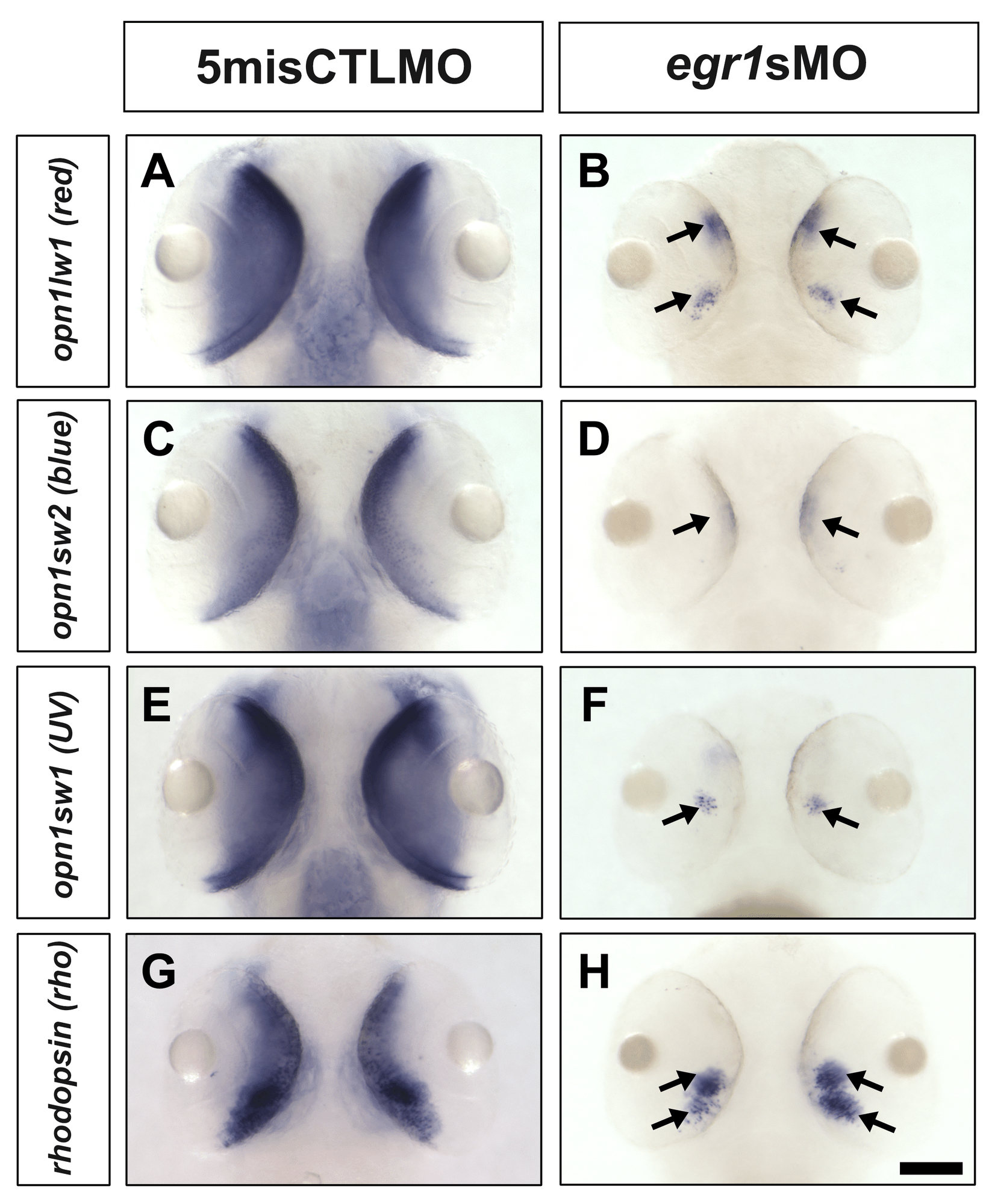
Fig. S2 In situ hybridization of opsinsat 72 hpf. In situ hybridization of opn1lw1 (red; A & B), opn1sw2 (blue; C & D), opn1sw1 (uv; E & F) and rhodopsin (rho; G & H) was conducted with the controls (5misCTLMO) and Egr1 morphants (egr1sMO) collected at 72 hpf. The staining of four opsins were strongly detected in the whole ONL of the control retinas (A, C, E and G), while their signal in the Egr1 morphants was restricted to the ventral patch and/or a few ONL cells (arrows in B, D, F and H). The ventral view of the embryos is shown in all pictures. To quantify the signal intensity of in situ hybridization, the number of embryos with a specific level of staining (Type 1 - ventral patch staining only, Type 2 – ventral patch staining plus some central PR layer staining, and Type 3 – ventral patch plus full PR layer staining) was counted and analyzed by Mann-Whitney test. The results show that there was a difference in the staining type between the controls and Egr1 morphants for all four opsins ([red opsin]: control counts (type 1–3): 0, 0, 12; Egr1-morphant counts: 5, 13, 0; U = 0, p-value < 0.001; [blue opsin]: control counts: 0, 0, 12; Egr1-morphant counts: 11, 7, 0; U = 0, p-value < 0.001; [uv opsin]: control counts: 0, 0, 12; Egr1-morphant counts: 13, 5, 1; U = 6, p-value < 0.001; [rho]: control counts: 0, 0, 9; Egr1-morphant counts: 15, 5, 0; U = 0, p-value < 0.001). In this figure, all controls are staining Type 3 while all Egr1 morphants are staining Type 2. Note that the effect of Egr1 knockdown on PR differentiation is likely caused by a delay in development, as the immunostaining of PR markers at 120 hpf shows that the differentiation of PRs in the Egr1 morphants was comparable to the controls (Figure 7). Scale bar = 100 µm.