
Fig. 7
- ID
- ZDB-IMAGE-130326-60
- Antibodies
- Publication
- Zhang et al., 2013 - The Role of egr1 in Early Zebrafish Retinogenesis
- All Figures
- Figures for Zhang et al., 2013
|
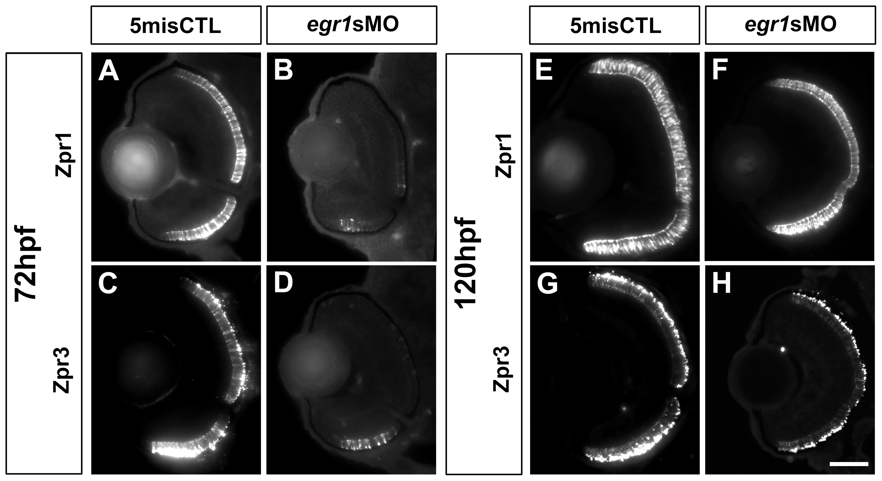
Fig. 7 PR differentiation was delayed in the Egr1-morphant retinas.
Immunohistochemical analysis of the PRs in the controls (5misCTLMO) and Egr1 morphants (egr1sMO) was performed with zpr1 (red-green double cones) and zpr3 (rods) at 72 hpf (A-D) and 120 (E-H) hpf. The signal of zpr1+ and zpr3+ cells was detected in the whole ONL of the controls at 72 hpf (A & C), while they were substantially reduced and restricted to a small region on the ventral ONL in the Egr1 morphants (B & D). Four staining types were defined as follows: Type 1: d ¼, 2: d ½, 3: d ¾, 4 = full retina. In these example images, the controls are staining Type 4 while the morphant images are staining Type 1. By 120 hpf, the differentiation of the zpr1+ and zpr3+ cells in the Egr1-morphant retinas (F & H) became more comparable to the controls (E & G). For all sections, the lens is on the left and dorsal is up. Scale bar = 50 µm.