
Fig. 4
- ID
- ZDB-IMAGE-130326-57
- Antibodies
- Publication
- Zhang et al., 2013 - The Role of egr1 in Early Zebrafish Retinogenesis
- All Figures
- Figures for Zhang et al., 2013
|
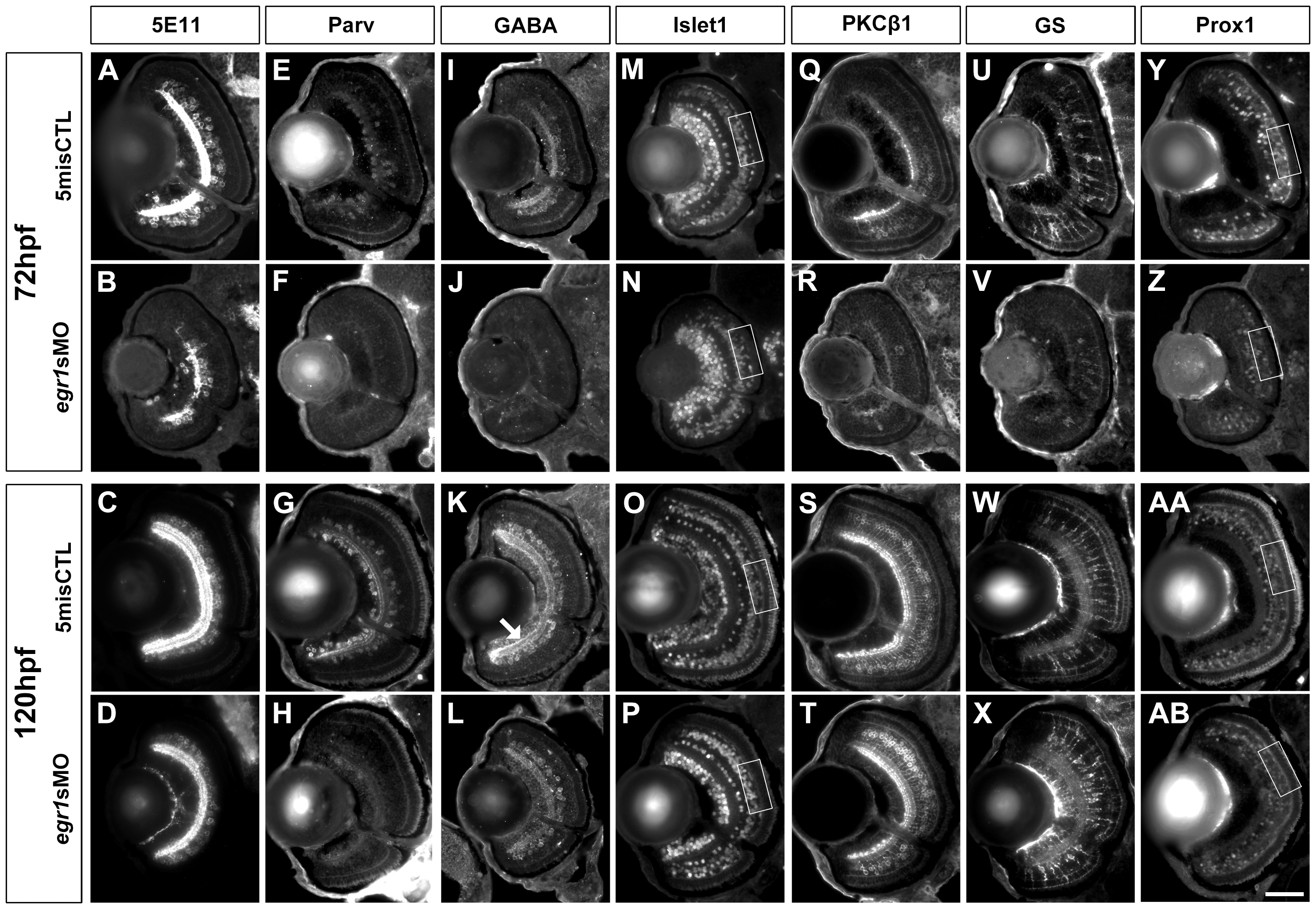
Fig. 4 Immunohistochemical analysis of the INL cells in the Egr1-morphant retinas.
Immunohistochemical analysis of the INL cells in the controls (5misCTLMO) and Egr1 morphants (egr1sMO) was performed with several cell markers at 72 and 120 hpf. These include anti-5E11 (5E11; A-D), anti-parvalbumin (Parv; E-H), anti-GABA (GABA; I-L) and anti-Islet1 (Islet1; M-P) for ACs; anti-PKC²1 (PKC; Q-T) for BCs; anti-GS (GS; U-X) for MCs; and Islet1 and anti-Prox1 (Prox1; Y-AB) for HCs. In short, the analysis has revealed that Egr1 knockdown specifically compromised the differentiation of Parv+ and GABA+ ACs. See text, Table 1 and 2 for further discussion and additional results for the specific effects on HCs differentiation in Figure 6. For all sections, the lens is on the left and dorsal is up. Scale bar = 50 µm.