
Fig. S1
- ID
- ZDB-IMAGE-130322-5
- Publication
- Choi et al., 2013 - In vivo monitoring of cardiomyocyte proliferation to identify chemical modifiers of heart regeneration
- All Figures
- Figures for Choi et al., 2013
|
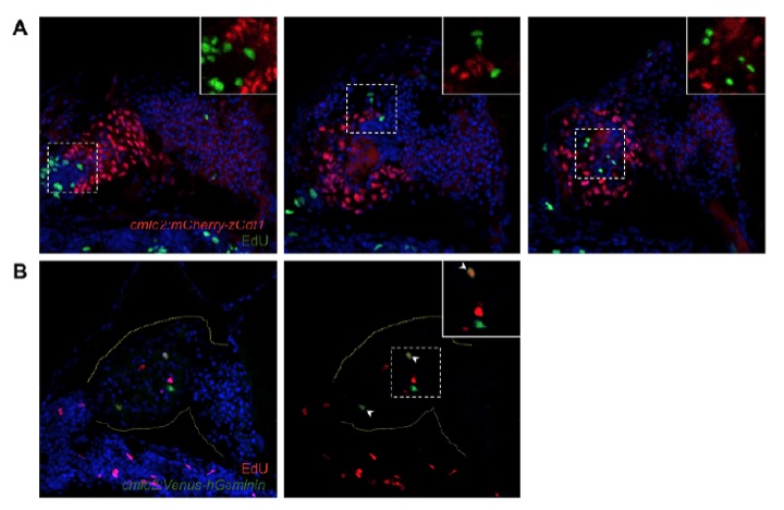
Fig. S1 Co-staining of cardiac FUCCI expression with EdU incorporation. (A) Cells expressing the cmlc2:mCherry-zCdt1 transgene (red) do not overlap with cells containing incorporated EdU (green). Images are representative maximum intensity projections from the same heart. DAPI, blue. Inset: High magnification of the boxed area. (B) A subset of cells expressing the cmlc2:Venus-hGeminin transgene (green) also contain incorporated EdU (red). DAPI, blue. Inset: High magnification of the boxed area. Arrowheads, co-expressing cells; dotted yellow line, outline of the heart. Note that many EdU+ cells in these images are not cardiomyocytes.