
Fig. 2
- ID
- ZDB-IMAGE-130320-2
- Antibodies
- Publication
- Sainath et al., 2013 - Plexin a3 and turnout regulate motor axonal branch morphogenesis in zebrafish
- All Figures
- Figures for Sainath et al., 2013
|
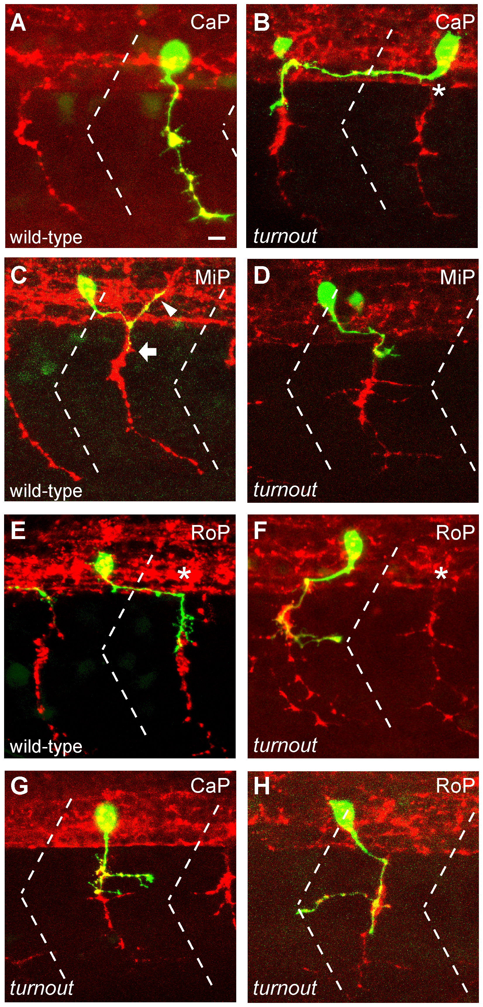
Fig. 2 turnout controls guidance and branching of primary motor axons.
(A, C, E) 24 hpf wild-type embryos injected with mnx1:GFP to visualize individual CaP, MiP and RoP motor neurons, and counter-stained with the SV2 antibody (red) to visualize the segmental exit points (asterisks). (B) A turnout CaP motor neuron projecting rostrally within the spinal cord, and exiting from an adjacent hemisegments midsegmental exit point. (D) A MiP motor neuron failing to migrate upon exit from the spinal cord. (F) A RoP motor axon projecting rostrally and exiting from an adjacent hemisegment exit. (G, H) turnout CaP and RoP motor neurons with correct axonal trajectories and ectopic branches. Dashed line indicates somite boundary. Scale bar, 10 µm.